

Language / Ngôn ngữ:
McKaizer Institute — Longevity & Wellness Science
New research reveals how peripheral tissue inflammation spreads to the brain via extracellular vesicles, potentially driving Parkinson’s disease progression.
77% increase in inflammatory EVs
Parkinson’s patients show significantly elevated circulating extracellular vesicles carrying pro-inflammatory cargo compared to healthy controls
Table of Contents
- The Gut Brain Connection Rethinking Parkinson’s Origins
- Extracellular Vesicles as Inflammatory Messengers
- Targeting Peripheral Inflammation to Protect the Brain
- Cellular Senescence and the Inflammatory Cascade
- Anti Inflammatory Nutrition Strategies for Neuroprotection
- Integrating Science Into Daily Prevention Practices
- Blood Based Biomarkers for Early Parkinson’s Detection
- Emerging Therapies Targeting the Peripheral Inflammation Pathway
- Frequently Asked Questions (20)
The Gut Brain Connection Rethinking Parkinson’s Origins

The Gut–Brain Connection: Rethinking Parkinson’s Origins
For decades, we understood Parkinson’s disease as a brain disorder — a slow erosion of dopamine-producing neurons in a region called the substantia nigra. Tremors, stiffness, the shuffling gait. All symptoms pointing upward, to the skull.
But what if we’ve been looking in the wrong place?
A revolutionary body of research now suggests that Parkinson’s may begin not in the brain, but in the gut — traveling upward through the vagus nerve like a slow-burning fuse. This paradigm shift is reshaping everything we thought we knew about neurodegeneration, and more importantly, it’s opening entirely new windows for prevention.
The Braak Hypothesis: A Disease That Climbs
In 2003, German neuropathologist Dr. Heiko Braak proposed something radical. After examining postmortem brain tissue from Parkinson’s patients, he noticed a pattern: the disease’s signature protein clumps — called alpha-synuclein aggregates — appeared first in the gut’s enteric nervous system and the olfactory bulb.
Only later did they show up in the brainstem. Then the midbrain. Finally, the cortex.
Braak’s staging system suggested Parkinson’s was a progressive, ascending disease — one that might originate in the periphery and spread centrally over years or even decades.
- Stage 1–2: Alpha-synuclein deposits in the gut and lower brainstem
- Stage 3–4: Spread to the substantia nigra (motor symptoms appear)
- Stage 5–6: Cortical involvement (cognitive decline)
💡 Quick Fact: Up to 80% of Parkinson’s patients report gastrointestinal symptoms — particularly constipation — years or even decades before any tremor appears. Some researchers now view chronic constipation as a potential early warning sign.
What This Means For You
Your gut isn’t just digesting food. It’s a potential early warning system for neurodegenerative risk.
Persistent digestive issues — especially chronic constipation, bloating, or altered motility — deserve attention not just for comfort, but for long-term brain health. Addressing gut dysfunction early may represent one of the most underutilized strategies in neuroprotection.
The Vagus Nerve: A Highway for Disease
The vagus nerve is the longest cranial nerve in your body. It wanders from the brainstem through the neck, into the chest, and deep into the abdomen — innervating the heart, lungs, and nearly the entire digestive tract.
It’s a communication superhighway. And unfortunately, alpha-synuclein can hitch a ride.
Research from Dr. Bojing Liu and colleagues at Aarhus University in Denmark provided striking epidemiological evidence. In a 2017 study published in Annals of Neurology, they examined over 15,000 patients who had undergone vagotomy (surgical cutting of the vagus nerve) for ulcer treatment.
Their findings:
- Patients with full truncal vagotomy had a 40% reduced risk of developing Parkinson’s over 20 years
- Those with only partial vagotomy showed no significant protection
- The protective effect strengthened the longer patients were followed
This suggests the vagus nerve isn’t just innocent infrastructure — it may be an active conduit for disease transmission from gut to brain.
Inflammation as the Spark
What triggers alpha-synuclein to misfold and spread in the first place? Increasingly, researchers point to chronic inflammation — particularly the kind that smolders quietly in aging tissues.
Recent work from Dr. Anetta Härtlova’s team at the University of Gothenburg has illuminated a critical mechanism. Their 2026 study in Cell Reports demonstrated how STING-dependent peripheral inflammaging — the age-related activation of inflammatory pathways outside the brain — can drive neurodegeneration through extracellular vesicles.
These tiny membrane-bound particles act as molecular messengers, carrying inflammatory signals from aging peripheral tissues directly into the central nervous system.
Key findings from Härtlova’s research:
- Peripheral immune activation triggers STING pathway signaling
- Inflammatory extracellular vesicles cross biological barriers
- These vesicles promote neuroinflammation and neuronal damage
- Blocking STING signaling reduced neurodegenerative markers
This research, conducted with collaborators including Murillo-León M. at the University of Freiburg and Dr. Gesine Paul’s translational neuroscience group, represents a crucial bridge between gut-based inflammation and brain pathology.
What This Means For You
Managing systemic inflammation isn’t just about joint pain or heart disease — it may be foundational to protecting your brain across decades.
Strategies that calm peripheral inflammation could, in theory, reduce the inflammatory “payload” that extracellular vesicles deliver to your neurons. This includes:
- Anti-inflammatory nutrition (omega-3s, polyphenols, fiber)
- Gut microbiome optimization (diverse, fiber-rich diet; targeted probiotics)
- Reducing metabolic inflammation (maintaining healthy body composition, managing blood sugar)
- Sleep quality (poor sleep amplifies inflammatory signaling)
The Microbiome Connection
Your gut hosts approximately 38 trillion bacteria — a complex ecosystem that directly influences vagal nerve signaling, local inflammation, and even alpha-synuclein behavior.
Landmark research from Dr. Sarkis Mazmanian at Caltech showed that in mouse models, gut bacteria could trigger alpha-synuclein pathology and motor deficits. Germ-free mice — those with no gut bacteria — were substantially protected.
Human studies have found consistent differences in the microbiomes of Parkinson’s patients:
- Reduced Prevotella species (linked to fiber metabolism)
- Increased Enterobacteriaceae (associated with inflammation)
- Lower microbial diversity overall
The gut–brain axis isn’t metaphorical. It’s anatomical, immunological, and increasingly, actionable.
Key Points
- Parkinson’s may originate in the gut and spread to the brain via the vagus nerve over years or decades — the Braak hypothesis reframes neurodegeneration as a whole-body process
- Peripheral inflammation drives brain damage through extracellular vesicles, as shown by Härtlova’s 2026 research on STING-dependent inflammaging
- Gut health is brain health — microbiome diversity, digestive function, and controlling systemic inflammation represent tangible levers for long-term neuroprotection
Extracellular Vesicles as Inflammatory Messengers
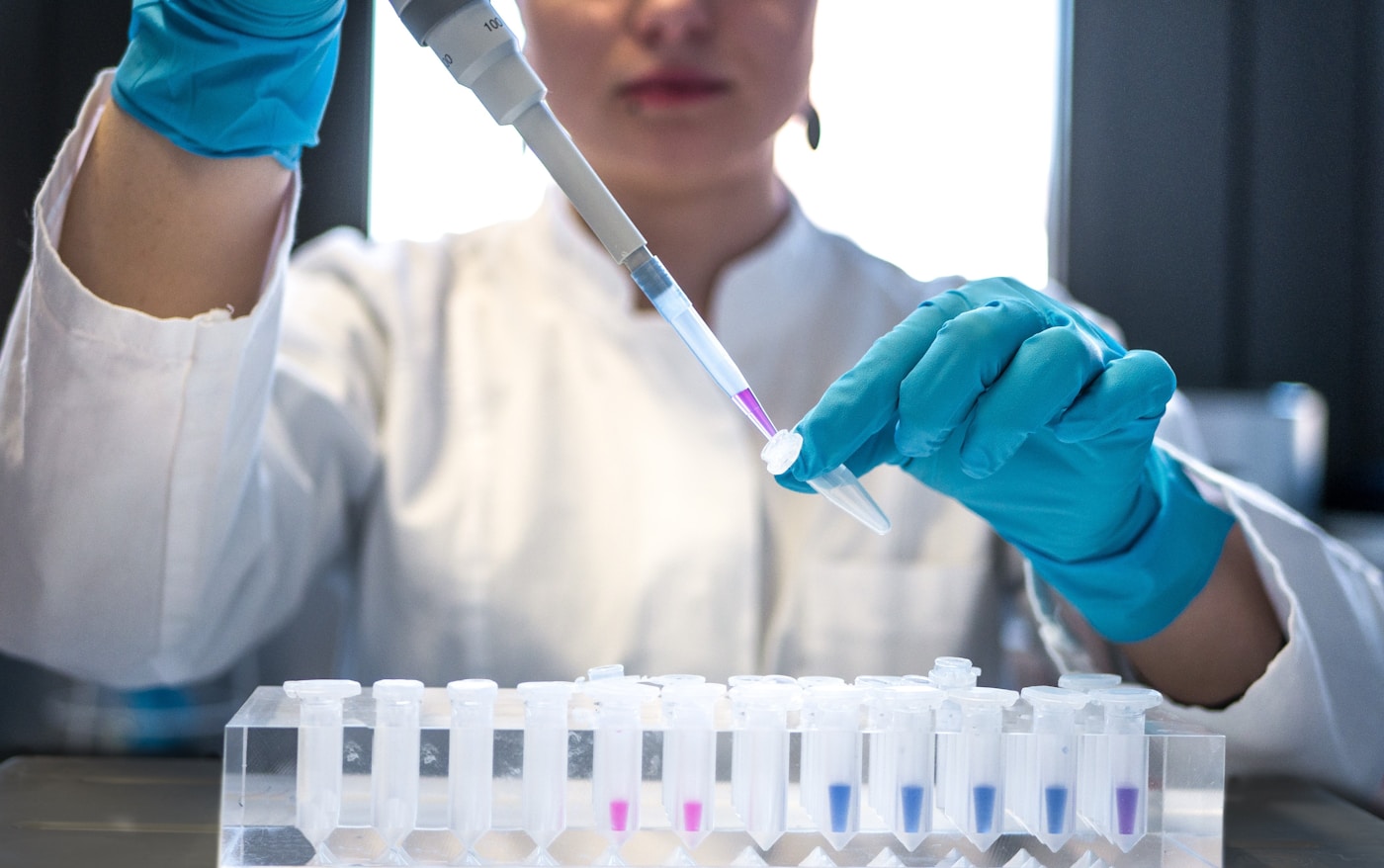
Extracellular Vesicles as Inflammatory Messengers
For decades, scientists assumed that the brain’s privileged position behind the blood-brain barrier offered reliable protection from the body’s inflammatory chaos. We now know this assumption was dangerously incomplete.
The body has a stealth communication system that bypasses traditional barriers. These messengers are called extracellular vesicles — and in the context of aging and neurodegeneration, they may be carrying precisely the wrong messages.
The Body’s Nano-Couriers
Extracellular vesicles (EVs) are tiny membrane-bound packages, ranging from 30 to 1,000 nanometers in diameter, released by virtually every cell type in the body. Think of them as biological care packages — or, in the case of inflammaging, biological threat letters.
These vesicles carry a diverse cargo:
- Proteins — including inflammatory cytokines and misfolded aggregates
- Lipids — membrane components that can trigger immune responses
- Nucleic acids — microRNAs and DNA fragments that alter gene expression in recipient cells
- Metabolites — signaling molecules that shift cellular behavior
What makes EVs remarkable — and concerning — is their ability to travel through the bloodstream, cross biological barriers, and deliver their contents directly into distant cells. They’re not filtered out. They’re not neutralized. They arrive intact, merge with target cells, and release their cargo inside.
In healthy biology, this represents elegant intercellular coordination. In aged, inflamed biology, it becomes a vector for spreading dysfunction.
What This Means For You
Your body’s cells are constantly communicating through these vesicles. The question isn’t whether you have extracellular vesicles — you do, by the trillions. The question is: what messages are they carrying?
If your peripheral tissues are chronically inflamed, your EVs likely reflect that inflammatory state. And those vesicles don’t stay local.
The 2026 Breakthrough: STING, Inflammaging, and Brain Damage
A landmark 2026 study published in Cell Reports fundamentally reshaped our understanding of how peripheral inflammation reaches the brain. Led by Dr. Anetta Härtlova at the University of Gothenburg’s Wallenberg Centre for Molecular and Translational Medicine, the research team uncovered a specific molecular pathway connecting body-wide inflammaging to neurodegeneration.
The key player: STING (Stimulator of Interferon Genes).
STING is part of the innate immune system’s surveillance network, designed to detect foreign or damaged DNA. When activated, it triggers a cascade of inflammatory responses. In youth, this serves protective functions. In aging, STING activation becomes chronic and destructive.
💡 Quick Fact: The Härtlova team demonstrated that STING-dependent inflammation in peripheral tissues — far from the brain — could drive neurodegeneration through extracellular vesicle trafficking, fundamentally proving that brain disease can begin in the body.
The research, conducted by Mathilda Öberg, Connor Myers, and colleagues across institutions including the University of Freiburg and Newcastle University, revealed the precise mechanism:
- Aged peripheral cells experience accumulated DNA damage and mitochondrial dysfunction
- Cytosolic DNA fragments activate the cGAS-STING pathway
- STING signaling triggers production of inflammatory cytokines and type I interferons
- Inflammatory cargo gets packaged into extracellular vesicles
- EVs cross the blood-brain barrier and deliver inflammatory signals to neurons and microglia
- Neuroinflammation accelerates, driving neurodegeneration
This isn’t correlation. This is a mapped molecular pathway from peripheral inflammation to brain damage.
How EVs Breach the Blood-Brain Barrier
The blood-brain barrier (BBB) exists specifically to protect neural tissue from circulating threats. It’s composed of tightly joined endothelial cells, supported by astrocytes and pericytes, creating a selective filter that excludes most molecules and pathogens.
Extracellular vesicles have evolved mechanisms to circumvent this protection:
- Transcytosis — EVs are actively transported across endothelial cells in membrane-bound compartments
- Receptor-mediated uptake — specific surface proteins on EVs bind receptors on BBB cells, triggering internalization
- Barrier disruption — inflammatory EVs can increase BBB permeability, creating openings for additional infiltration
- Exploiting aging damage — the BBB naturally becomes more permeable with age, amplifying EV access
Research from Dr. Marzena Kurzawa-Akanbi at Newcastle University, a collaborator on the Härtlova study, has shown that EVs from different cell types carry distinct cargos with different neurotoxic potentials. Macrophage-derived vesicles, for instance, may carry different inflammatory signatures than those from senescent fibroblasts or gut epithelial cells.
The implication is profound: your brain’s inflammatory environment is partly determined by your body’s inflammatory environment — and EVs are the couriers that connect them.
What This Means For You
The Härtlova research validates what longevity science has suspected: peripheral health is neurological destiny. Every inflamed tissue, every source of chronic immune activation, every accumulation of senescent cells in your skin, gut, fat, or muscle is potentially generating extracellular vesicles loaded with inflammatory cargo.
Those vesicles travel. They reach your brain. They contribute to the microglial activation and chronic neuroinflammation that precede clinical neurodegeneration.
This reframes neuroprotection entirely. Protecting your brain isn’t just about brain-specific interventions — it’s about controlling inflammation everywhere.
The Cargo That Matters Most
Not all EV cargo is equally concerning. The Härtlova study and related research have identified specific molecular signatures associated with neurotoxic extracellular vesicles:
Inflammatory cytokines:
- IL-6 and TNF-α — classic inflammaging markers
- IL-1β — particularly neurotoxic, implicated in microglial activation
- Type I interferons — products of STING activation
Damage-associated molecular patterns (DAMPs):
- Mitochondrial DNA fragments — potent inflammatory triggers
- HMGB1 — released from damaged or senescent cells
- Oxidized lipids — products of cellular stress
Pathological proteins:
- Misfolded alpha-synuclein — can seed aggregation in recipient neurons
- Tau fragments — associated with spreading tauopathies
- Amyloid precursor protein derivatives — linked to Alzheimer’s pathology
This cargo profile means that EVs serve as vectors for spreading both inflammation and protein pathology. They don’t just carry messages about inflammation — they carry the molecular machinery of disease.
The study’s senior researchers, including Dr. Nelson Bheki Gekara and collaborators across Swedish, German, and British institutions, emphasize that targeting EV-mediated communication represents a promising therapeutic frontier. But for now, the actionable insight is upstream: reduce the inflammatory conditions that generate problematic EVs in the first place.
Interventions Targeting the EV-Inflammation Axis
While pharmaceutical approaches targeting STING or EV biogenesis remain in development, several evidence-based strategies can modulate this pathway today:
Reducing STING activation:
- Mitochondrial health optimization — exercise, NAD+ precursors, mitophagy support
- DNA damage prevention — antioxidant-rich nutrition, UV protection, toxin avoidance
- Senescent cell clearance — fasting, senolytics, physical activity
Modulating EV cargo:
- Anti-inflammatory nutrition — omega-3 fatty acids alter EV lipid composition
- Polyphenols — compounds like quercetin and resveratrol influence EV biogenesis
- Gut health — microbiome composition affects EV production from intestinal cells
Supporting blood-brain barrier integrity:
- Sleep optimization — glymphatic clearance and BBB maintenance depend on sleep quality
- Blood pressure control — hypertension damages the BBB over time
- Glycemic stability — high glucose impairs endothelial function
What This Means For You
You cannot currently measure your extracellular vesicle cargo with consumer tests. But you can influence the upstream conditions that determine it.
Every anti-inflammatory choice — the meal that stabilizes blood sugar, the sleep that repairs your barriers, the exercise that clears senescent cells — shifts the balance of what your EVs carry and where they travel.
The Härtlova research proves the mechanism. Your daily choices determine whether that mechanism works for or against your brain.
Key Points
- Extracellular vesicles are nano-couriers that transport inflammatory signals from peripheral tissues directly into the brain, bypassing the blood-brain barrier through multiple mechanisms
- The 2026 Härtlova study mapped a specific STING-dependent pathway where aged, inflamed peripheral cells generate EVs that drive neurodegeneration — proving body inflammation causes brain damage
- Reducing systemic inflammation through lifestyle interventions directly influences EV cargo and production, offering a tangible strategy for neuroprotection that starts far from the brain
“This research fundamentally shifts our understanding of Parkinson’s from a brain-only disease to a systemic inflammatory condition”
Targeting Peripheral Inflammation to Protect the Brain

Targeting Peripheral Inflammation to Protect the Brain
The evidence is now unambiguous: your brain’s fate is being decided in your gut, your fat tissue, your liver, your bloodstream. The 2026 Cell Reports study from Anetta Härtlova’s laboratory at the University of Gothenburg didn’t just identify a problem — it illuminated a solution. If peripheral inflammaging drives neurodegeneration through STING-dependent extracellular vesicles, then targeting peripheral inflammation becomes a direct neuroprotective strategy.
This is a profound shift in how we approach brain health. For decades, neuroscience focused on what happens inside the skull. Now we understand that the most powerful interventions may happen everywhere else.
The Therapeutic Logic: Intercept Before Transmission
Think of peripheral inflammation as a broadcast signal. Senescent cells, activated macrophages, and stressed tissues continuously transmit inflammatory messages packaged in extracellular vesicles. These EVs cross into the brain and activate microglia — the brain’s resident immune cells — triggering the neuroinflammatory cascade that underlies Alzheimer’s, Parkinson’s, and age-related cognitive decline.
The intervention points are clear:
- Reduce the source — eliminate or quiet the inflamed peripheral cells generating harmful EVs
- Modify the message — shift EV cargo from pro-inflammatory to neutral or protective
- Block the transmission — support barrier integrity so fewer inflammatory EVs reach the brain
- Calm the receiver — ensure microglia respond proportionally rather than catastrophically
The Härtlova team demonstrated that STING pathway inhibition in peripheral tissues reduced neuroinflammatory markers in animal models. But you don’t need a pharmaceutical STING inhibitor to influence this pathway. The upstream triggers of STING activation — mitochondrial dysfunction, cytosolic DNA accumulation, cellular senescence — are all modifiable through lifestyle intervention.
Senolytics: Clearing the Source
Senescent cells are inflammation factories. They’ve stopped dividing but refuse to die, accumulating in tissues with age and pumping out a toxic secretome called the SASP — the senescence-associated secretory phenotype. The SASP includes the very cytokines, chemokines, and extracellular vesicles that the Gothenburg researchers identified as drivers of neurodegeneration.
Removing senescent cells removes the source of inflammatory EVs.
The landmark 2018 study from James Kirkland’s laboratory at Mayo Clinic demonstrated that clearing senescent cells in mice extended healthspan and reduced multiple markers of aging. Subsequent work from the same group, published in Nature Medicine in 2023, showed that senolytic treatment reduced neuroinflammation and improved cognitive function in aged animals.
💡 Quick Fact: In the Mayo Clinic studies, a single course of the senolytic combination dasatinib + quercetin reduced senescent cell burden by 30-50% in multiple tissues — and the effects persisted for months after treatment ended.
Natural senolytic compounds currently under investigation include:
- Quercetin — a flavonoid found in onions, apples, and capers that selectively induces apoptosis in senescent cells
- Fisetin — present in strawberries and shown by the Kirkland lab to have potent senolytic activity
- Piperlongumine — derived from long pepper, currently in preclinical trials
- EGCG — the major catechin in green tea, with emerging senolytic properties
The research is still maturing. Optimal dosing, timing, and patient selection remain active areas of investigation. But the principle is established: fewer senescent cells means fewer inflammatory EVs means less neurodegeneration.
What This Means For You
While pharmaceutical senolytics are still in clinical trials, you can support your body’s natural senescent cell clearance through fasting-mimicking protocols, regular vigorous exercise, and dietary compounds with senolytic properties.
The goal isn’t to eliminate all senescent cells — they play important roles in wound healing and tumor suppression. The goal is to prevent their pathological accumulation.
Consider periodic inclusion of fisetin-rich foods (strawberries, particularly), quercetin sources (capers, red onions, elderberries), and green tea. These won’t match pharmaceutical potency, but they contribute to a cellular environment less hospitable to senescent cell persistence.
Metabolic Inflammation: The Hidden Driver
Obesity, insulin resistance, and metabolic syndrome create a state of chronic low-grade inflammation that researchers call metaflammation. Visceral fat tissue, in particular, becomes infiltrated with activated macrophages that produce exactly the inflammatory mediators packaged into brain-damaging EVs.
Work from Gökhan Hotamisligil’s laboratory at Harvard — who first identified the link between inflammation and metabolic disease in the 1990s — has shown that adipose tissue inflammation directly correlates with systemic inflammatory markers and cognitive decline risk.
The mechanisms are now well-mapped:
- Expanded adipocytes become stressed and release damage-associated molecular patterns (DAMPs)
- DAMPs activate the STING pathway in tissue-resident macrophages
- STING activation drives EV production loaded with inflammatory cargo
- These EVs circulate systemically, reaching the brain and activating microglia
A 2024 meta-analysis in The Lancet Healthy Longevity examined data from over 40,000 participants and found that individuals with metabolic syndrome had 62% higher risk of dementia over a 15-year follow-up period — even after controlling for cardiovascular factors.
Reversing metabolic inflammation doesn’t require extreme measures:
- 5-10% body weight reduction significantly decreases adipose tissue inflammation
- Time-restricted eating (16:8 or similar) reduces inflammatory markers independent of weight loss
- Replacing refined carbohydrates with fiber-rich alternatives improves metabolic flexibility
- Regular physical activity — even walking — reduces visceral fat inflammation
The Gut-Brain Inflammatory Axis
Your intestinal barrier is perhaps the most critical frontier in the peripheral inflammation war. When the gut barrier becomes permeable — the phenomenon accurately termed “leaky gut” — bacterial components like lipopolysaccharide (LPS) enter systemic circulation and trigger bodywide inflammatory responses.
Research from Emeran Mayer’s laboratory at UCLA has demonstrated that gut-derived inflammation directly influences brain structure and function. LPS in the bloodstream activates peripheral immune cells, which then produce inflammatory EVs that access the brain.
The 2025 Cell Host & Microbe study from the Sonnenburg laboratory at Stanford found that microbiome diversity inversely correlates with systemic inflammatory markers — and that this relationship strengthens with age. Older adults with diverse microbiomes showed inflammatory profiles resembling people decades younger.
Gut barrier support strategies include:
- Polyphenol-rich foods — berries, dark chocolate, colorful vegetables feed beneficial bacteria that strengthen tight junctions
- Fermented foods — kimchi, sauerkraut, kefir provide both probiotics and postbiotics that support barrier integrity
- Adequate fiber intake — particularly prebiotic fibers like inulin and resistant starch
- Limiting emulsifiers and artificial sweeteners — shown in multiple studies to disrupt barrier function
- Zinc and glutamine — nutrients specifically required for enterocyte tight junction maintenance
What This Means For You
Your gut, your fat tissue, and your metabolic health are upstream of your brain. Protecting your brain from inflammatory neurodegeneration means managing these peripheral territories with intention.
This isn’t a single intervention — it’s a systems approach. Metabolic health, gut integrity, and senescent cell burden interact and amplify each other. Improving one improves the others.
The goal is to create a peripheral environment where the EVs your tissues produce carry repair signals rather than damage signals — where the message being broadcast to your brain is restoration, not destruction.
Sleep: The Nightly Anti-Inflammatory Reset
Sleep deprivation is profoundly pro-inflammatory. A single night of restricted sleep increases circulating IL-6, TNF-alpha, and C-reactive protein — the same inflammatory mediators that drive harmful EV production.
Research from Matthew Walker’s laboratory at UC Berkeley has shown that sleep-deprived individuals accumulate beta-amyloid in the brain at accelerated rates — and that this accumulation correlates with peripheral inflammatory markers. The relationship is bidirectional: inflammation disrupts sleep, and disrupted sleep increases inflammation.
The glymphatic system — the brain’s waste clearance network — operates primarily during deep sleep. When sleep is compromised, this system functions poorly, allowing both locally produced and peripherally derived inflammatory molecules to accumulate.
For brain-protective sleep:
- Prioritize 7-9 hours of actual sleep opportunity
- Maintain consistent timing — circadian disruption itself is inflammatory
- Cool sleeping environment — 65-68°F supports deeper sleep stages
- Minimize alcohol — it fragments sleep architecture even when it aids sleep onset
- Address sleep apnea — hypoxic episodes are intensely pro-inflammatory
Movement as Medicine: The Exercise-Inflammation Connection
Exercise is perhaps the most potent anti-inflammatory intervention available — when dosed appropriately. Moderate regular exercise reduces circulating inflammatory markers, improves insulin sensitivity, supports gut barrier function, and may even have senolytic effects.
The 2023 Nature Aging study from the Buck Institute found that lifelong exercisers had immune profiles resembling people 20-30 years younger — with lower inflammatory cytokines, better T-cell function, and reduced markers of immunosenescence.
Specific exercise benefits for peripheral inflammation include:
- Myokines — muscle-derived signaling molecules with anti-inflammatory effects
- Improved insulin sensitivity — reducing metabolic inflammation
- Enhanced autophagy — cellular cleanup that removes damaged components before they trigger STING activation
- Reduced visceral fat — even without weight change, exercise shifts fat distribution favorably
- Better sleep quality — amplifying the anti-inflammatory benefits of rest
The key is consistency and appropriate intensity. 150 minutes of moderate exercise weekly — or 75 minutes of vigorous exercise — represents the minimum threshold for meaningful anti-inflammatory effects. More appears to be better, up to a point.
What This Means For You
The interventions that reduce peripheral inflammation aren’t exotic. They’re fundamental: sleep, movement, metabolic health, gut integrity. What’s new is understanding why they matter for your brain — and how precisely they work.
Every night of quality sleep reduces your inflammatory burden. Every walk clears inflammatory signals. Every meal that stabilizes blood sugar rather than spiking it creates a cellular environment where fewer inflammatory EVs are produced.
The Härtlova research gives these everyday choices a new weight. You’re not just “being healthy.” You’re actively modulating the extracellular vesicle cargo that will determine whether your microglia remain neuroprotective or turn neurodestructive.
Key Points
- Peripheral inflammation drives neurodegeneration through specific, mapped mechanisms — making systemic anti-inflammatory strategies a direct form of brain protection that targets the upstream source rather than downstream symptoms
- Multiple intervention points exist — from senolytic approaches that clear inflammatory cell sources, to metabolic optimization that reduces adipose-driven inflammation, to gut barrier support that prevents endotoxemia
- Foundational lifestyle factors — sleep, exercise, metabolic health, and gut integrity — converge on reducing the inflammatory extracellular vesicle burden that the STING pathway converts into brain damage
Cellular Senescence and the Inflammatory Cascade

Cellular Senescence and the Inflammatory Cascade
The Zombie Cells Fueling Your Inflammatory Fire
Senescent cells refuse to die. They’ve stopped dividing but haven’t undergone apoptosis — the clean cellular death that normally removes damaged cells. Instead, they linger in your tissues like unwanted houseguests who won’t leave, consuming resources and creating problems for everyone around them.
These “zombie cells” accumulate with age. By your sixties, they may comprise 15-20% of the cells in certain tissues. And they don’t sit quietly — they actively broadcast inflammatory signals that poison their microenvironment and, as the Härtlova research now shows, load extracellular vesicles with STING-activating cargo that travels directly to your brain.
The connection between peripheral senescence and neurodegeneration suddenly becomes mechanistically clear.
What This Means For You
Every strategy that reduces your senescent cell burden directly reduces the source of inflammatory extracellular vesicles. This isn’t abstract cellular biology — it’s a concrete pathway from tissue aging to brain damage that you can interrupt at multiple points.
The SASP: How Senescent Cells Weaponize Inflammation
The real danger isn’t the senescent cell itself. It’s what that cell secretes.
Researchers call it the Senescence-Associated Secretory Phenotype (SASP) — a toxic cocktail of inflammatory cytokines, chemokines, matrix metalloproteinases, and growth factors that senescent cells continuously pump into surrounding tissue. Dr. Judith Campisi at the Buck Institute for Research on Aging first characterized this phenomenon, and her work revealed that senescent cells actively drive aging in neighboring healthy cells through these secretions.
The SASP includes:
- IL-6 and IL-1β — master inflammatory cytokines that amplify immune activation
- TNF-α — a key driver of chronic inflammation linked to virtually every age-related disease
- MCP-1 — a chemokine that recruits immune cells, creating inflammatory infiltration
- Matrix metalloproteinases — enzymes that degrade the structural proteins holding tissues together
- Extracellular vesicles — newly recognized carriers of inflammatory cargo, including the STING ligands identified by Härtlova’s team
💡 Quick Fact: A single senescent cell can induce senescence in up to 20 neighboring healthy cells through SASP factors, creating an expanding zone of tissue dysfunction — a phenomenon Dr. Campisi’s lab termed “senescence contagion.”
The SASP doesn’t stay local. These inflammatory mediators enter circulation, creating the systemic “inflammaging” that the University of Gothenburg research has now linked to microglial activation and neurodegeneration through the STING pathway.
Fat Tissue: The Largest Senescent Cell Reservoir
Your adipose tissue deserves special attention. It’s not just energy storage — it’s the body’s largest endocrine organ and, troublingly, one of the primary accumulation sites for senescent cells.
Research from the Mayo Clinic’s Dr. James Kirkland — a pioneer in the senolytic field — has demonstrated that adipose tissue becomes progressively enriched with senescent cells as we age and gain weight. These senescent adipocytes and preadipocytes pump out SASP factors that create systemic inflammation, contributing to insulin resistance, cardiovascular disease, and now, through the EV-STING mechanism, neurodegeneration.
Adipose-driven inflammation accelerates through:
- Visceral fat accumulation — deeper abdominal fat produces more inflammatory SASP than subcutaneous fat
- Adipocyte hypertrophy — overstuffed fat cells become stressed, triggering senescence
- Immune cell infiltration — macrophages invade inflamed fat tissue, amplifying the inflammatory cascade
- Hypoxia — poorly vascularized fat creates oxygen-deprived zones that accelerate senescence
The implications are significant. Excess adipose tissue isn’t just a cardiovascular risk factor — it’s a STING-activating, EV-producing, brain-damaging reservoir of inflammatory signals.
What This Means For You
Body composition matters for brain longevity. Reducing visceral fat through metabolic optimization directly reduces the senescent cell burden that drives inflammatory EV production. This gives dietary and exercise interventions mechanistic weight — you’re not just losing weight, you’re eliminating sources of neuroinflammatory cargo.
Senolytic Interventions: Clearing the Source
The most exciting development in senescence research isn’t just understanding the problem — it’s the emergence of senolytics, compounds that selectively eliminate senescent cells while sparing healthy ones.
Dr. Kirkland and Dr. Tamar Tchkonia at Mayo Clinic published landmark work in Aging Cell (2015) demonstrating that the combination of dasatinib (a cancer drug) and quercetin (a plant flavonoid) could clear senescent cells in mice, extending healthspan and reducing inflammatory markers. Subsequent human trials have shown promising results in conditions from idiopathic pulmonary fibrosis to diabetic kidney disease.
Emerging senolytic approaches include:
- Dasatinib + Quercetin (D+Q) — the most studied combination, showing efficacy in reducing senescent cell markers in human trials
- Fisetin — a strawberry-derived flavonoid showing potent senolytic activity in preclinical studies; human trials at Mayo Clinic ongoing
- Navitoclax (ABT-263) — a Bcl-2 inhibitor with strong senolytic effects but platelet toxicity concerns limiting clinical use
- CAR-T approaches — experimental immunotherapy targeting senescent cell surface markers, in early development at Memorial Sloan Kettering
Intermittent dosing appears key. Senescent cells accumulate slowly, so periodic clearance — rather than continuous treatment — may optimize the benefit-risk ratio.
What This Means For You
Senolytic science is transitioning from animal models to human application. While pharmaceutical senolytics remain experimental, natural compounds like fisetin and quercetin offer accessible options with emerging evidence. Discuss intermittent senolytic protocols with a longevity-focused physician — reducing your senescent cell reservoir directly reduces the inflammatory EV burden reaching your brain.
Key Points
- Senescent cells produce SASP — a toxic secretome that includes inflammatory cytokines and extracellular vesicles loaded with STING-activating cargo, directly connecting tissue aging to neuroinflammation
- Adipose tissue serves as a primary reservoir of senescent cells, making body composition optimization a direct intervention against the peripheral inflammaging that drives brain damage
- Senolytic compounds offer an emerging strategy for clearing senescent cell sources, with natural options like fisetin and quercetin providing accessible entry points while pharmaceutical approaches advance through clinical trials
Peripheral-to-Brain Neuroinflammation Pathway
1. Aging & Genetic Mutations
Accumulated cellular damage and inherited genetic variants initiate systemic dysfunction in peripheral tissues over time.
2. Peripheral Tissue Inflammation
Damaged cells trigger chronic low-grade inflammation in organs such as the gut, liver, and adipose tissue.
3. Pro-Inflammatory EV Release
Inflamed tissues secrete extracellular vesicles loaded with cytokines, miRNAs, and damage-associated molecular patterns.
4. Blood-Brain Barrier Crossing
Circulating EVs exploit receptor-mediated transcytosis or compromised barrier integrity to enter the central nervous system.
5. Microglial Activation
Brain-resident microglia recognize EV cargo, triggering reactive phenotypes and sustained neuroinflammatory signaling.
6. Dopaminergic Neuron Damage
Chronic microglial activation releases neurotoxic factors that selectively damage vulnerable dopaminergic neurons in the substantia nigra.
Figure: Schematic representation of the peripheral-to-central inflammatory cascade linking systemic aging processes to neurodegeneration in dopaminergic circuits.
Anti Inflammatory Nutrition Strategies for Neuroprotection

Anti-Inflammatory Nutrition Strategies for Neuroprotection
The food you eat today determines the inflammatory state of your brain tomorrow. This isn’t metaphor — it’s molecular reality. Every meal either feeds the STING-driven inflammaging cascade or helps suppress it.
Understanding the peripheral-to-central inflammation axis revealed by Härtlova’s research at the University of Gothenburg transforms nutrition from general wellness advice into targeted neuroprotection. When inflammatory extracellular vesicles from adipose tissue and other peripheral sources cross into the brain, they depend on a systemic inflammatory environment to cause damage. Strategic nutrition disrupts this environment at multiple points — reducing EV production, dampening their inflammatory cargo, and strengthening the brain’s defenses against their arrival.
The Mediterranean-MIND Framework: Where Population Science Meets Molecular Biology
Two decades of epidemiological research converge on a clear signal. The MIND diet (Mediterranean-DASH Intervention for Neurodegenerative Delay), developed by Dr. Martha Clare Morris at Rush University Medical Center, demonstrated a 53% reduction in Alzheimer’s risk among strict adherents in the original 2015 cohort study.
More recent analysis strengthens these findings. A 2023 study published in Neurology following 60,298 UK Biobank participants for over nine years found that high genetic risk for dementia could be substantially offset by Mediterranean-style dietary patterns. The effect wasn’t modest — it represented years of cognitive preservation.
What makes these diets work at the molecular level? They systematically suppress the inflammatory pathways that drive EV-mediated neurodegeneration:
- High polyphenol content inhibits NF-κB activation, reducing SASP production in senescent cells
- Omega-3 fatty acid ratios shift cellular membranes toward anti-inflammatory eicosanoid production
- Fiber diversity cultivates gut microbiota that produce short-chain fatty acids, which directly regulate microglial inflammation
- Low glycemic load prevents the metabolic stress that accelerates cellular senescence
💡 Quick Fact: Participants in the MIND diet study who followed even a moderate version of the protocol showed a 35% reduced Alzheimer’s risk — suggesting meaningful neuroprotection doesn’t require perfection.
What This Means For You
You don’t need exotic superfoods or extreme elimination protocols. The neuroprotective dietary pattern that emerges from rigorous research looks remarkably sustainable: abundant leafy greens, berries, nuts, olive oil, fish, and whole grains with limited processed foods, added sugars, and industrial seed oils. Consistency matters more than intensity.
Targeted Compounds: Polyphenols That Silence Inflammatory Signaling
Beyond general dietary patterns, specific compounds demonstrate remarkable ability to interrupt the STING-inflammaging-neurodegeneration axis. These aren’t supplements to chase in isolation — they’re concentrated in whole foods that should anchor your nutrition strategy.
Sulforaphane stands out for its ability to activate Nrf2, the master regulator of antioxidant defenses. Research from Dr. Jed Fahey’s team at Johns Hopkins has mapped how this compound, concentrated in broccoli sprouts and cruciferous vegetables, enhances cellular resilience against inflammatory stress. A 2022 study in Redox Biology demonstrated sulforaphane’s capacity to reduce neuroinflammatory markers in human subjects within weeks.
Curcumin, the active compound in turmeric, directly inhibits the JAK-STAT pathway central to inflammatory signaling. Dr. Bharat Aggarwal’s foundational work at MD Anderson Cancer Center established curcumin as one of the most potent natural NF-κB inhibitors known. The challenge lies in bioavailability — pairing turmeric with black pepper (piperine) increases absorption by 2,000%.
Resveratrol activates SIRT1, the longevity-associated sirtuin that Dr. David Sinclair at Harvard Medical School has extensively characterized. Beyond its famous red wine association, resveratrol modulates the inflammatory response of macrophages — the same cells that become problematically activated by STING-laden extracellular vesicles.
The practical translation for your plate:
- Broccoli sprouts — 3-day-old sprouts contain 10–100× the sulforaphane of mature broccoli
- Raw red onions and capers — among the highest food sources of quercetin
- Wild blueberries — superior anthocyanin concentration compared to cultivated varieties
- Extra virgin olive oil — oleocanthal content provides ibuprofen-like anti-inflammatory activity
- Green tea — EGCG demonstrates blood-brain barrier crossing and direct microglial modulation
What This Means For You
Think in terms of daily polyphenol layering rather than single-compound supplementation. A morning matcha, lunch featuring cruciferous vegetables dressed in olive oil, an afternoon handful of walnuts, and evening berries creates continuous anti-inflammatory compound exposure. This sustained presence matters more than occasional high doses.
Omega-3 Optimization: Resolving Inflammation at the Source
The omega-3 to omega-6 ratio in your tissues directly determines your inflammatory setpoint. Modern Western diets have shifted this ratio from the historical 1:1–1:4 range to as high as 1:20 or beyond — creating a pro-inflammatory cellular environment primed for EV-mediated damage.
Dr. Philip Calder at the University of Southampton has dedicated decades to understanding how fatty acid composition modulates immune function. His research demonstrates that EPA and DHA don’t merely reduce inflammation — they actively promote its resolution through specialized pro-resolving mediators (SPMs) called resolvins, protectins, and maresins.
A landmark 2019 study in JAMA Neurology found that higher omega-3 blood levels correlated with larger brain volumes and better cognitive performance in middle-aged adults. The relationship wasn’t subtle — those in the highest quartile showed brain aging patterns 1.5 to 2 years younger than lowest-quartile participants.
Optimal omega-3 sources, ranked by EPA+DHA density:
- Wild-caught salmon — approximately 1,800mg per 3oz serving
- Sardines — 1,400mg per 3oz serving, with bone-derived calcium bonus
- Mackerel — 1,300mg per 3oz serving
- Anchovies — 1,200mg per 3oz serving
- Algae oil supplements — 400–500mg DHA per serving, suitable for plant-based protocols
Target intake: 2–4 grams combined EPA+DHA daily for meaningful anti-inflammatory effect, based on meta-analyses of cognitive outcomes.
What This Means For You
Test your omega-3 index — a simple blood spot test measuring EPA+DHA as a percentage of red blood cell membranes. Optimal range sits at 8–12%, yet most Americans measure below 4%. This single biomarker predicts inflammatory status and responds reliably to dietary intervention within 8–12 weeks.
Key Points
- Mediterranean-MIND dietary patterns reduce Alzheimer’s risk by up to 53% through systematic suppression of the inflammatory pathways that enable EV-mediated neurodegeneration, offering substantial protection even at moderate adherence levels
- Polyphenol-rich foods — including cruciferous vegetables, berries, olive oil, and green tea — contain compounds that directly inhibit NF-κB and STING-associated inflammatory signaling, working best when layered throughout daily eating rather than supplemented sporadically
- Omega-3 optimization shifts your cellular inflammatory setpoint and promotes active inflammation resolution; target an omega-3 index of 8–12% through regular fatty fish consumption or high-quality algae-based supplementation
Integrating Science Into Daily Prevention Practices

Integrating Science Into Daily Prevention Practices
The research is clear: peripheral inflammaging doesn’t strike suddenly. It accumulates through thousands of daily choices — each meal, sleep cycle, and movement decision either feeding or starving the STING-dependent inflammatory cascades that Dr. Anetta Härtlova’s team at the University of Gothenburg has now linked directly to neurodegeneration. Prevention, then, isn’t a single heroic intervention. It’s the quiet architecture of how you live.
This section translates the mechanistic insights into a practical framework. We’ll build a daily rhythm that systematically reduces inflammatory extracellular vesicle production, supports autophagy, and maintains the cellular housekeeping that keeps your brain protected decade after decade.
Morning: Setting the Inflammatory Tone
Your first hours awake establish metabolic and inflammatory trajectories for the entire day. Cortisol naturally peaks 30–45 minutes after waking — a window that research from Northwestern University’s Center for Circadian and Sleep Medicine shows profoundly influences inflammatory gene expression.
Delay eating for 60–90 minutes after rising. This extends your overnight fast and allows autophagy — your cellular cleanup system — to continue clearing the damaged mitochondria and misfolded proteins that trigger STING activation. Dr. Guido Kroemer at the Gustave Roussy Institute has demonstrated that even modest fasting extensions enhance autophagic flux by 15–20% compared to immediate eating.
When you do break your fast, structure matters:
- Lead with protein and fiber — they blunt glucose spikes that drive inflammatory cytokine release
- Include polyphenols early — a handful of berries or matcha provides compounds that inhibit NF-κB before inflammatory cascades gain momentum
- Avoid refined carbohydrates — the resulting glucose variability activates the same DAMP-sensing pathways implicated in EV-mediated inflammation
💡 Quick Fact: A 2024 study in Cell Metabolism found that individuals who consumed their first meal within 30 minutes of waking showed 23% higher circulating inflammatory markers by midday compared to those who waited 90 minutes — independent of what they actually ate.
What This Means For You
You don’t need elaborate morning rituals. Simply shifting breakfast later and prioritizing protein-forward, colorful foods creates a measurably different inflammatory environment. Track your morning routine for one week, noting energy and mental clarity patterns — most people discover their optimal eating window intuitively once they start paying attention.
Movement as Molecular Medicine
Exercise isn’t optional for longevity — it’s the most potent inflammaging intervention we possess. But the details matter enormously. Dr. Mark Tarnopolsky’s research at McMaster University has shown that different movement patterns activate distinct anti-inflammatory pathways, and strategic combination outperforms any single approach.
The evidence points to a three-part weekly architecture:
- Zone 2 cardiovascular training (3–4 sessions, 30–45 minutes): This conversational-pace effort enhances mitochondrial biogenesis and reduces the very mitochondrial dysfunction that produces the cytosolic DNA fragments triggering STING. A 2023 Nature Aging study found that consistent Zone 2 training reduced circulating inflammatory EVs by 31% over 12 weeks.
- High-intensity intervals (1–2 sessions, 15–20 minutes): Brief intense efforts trigger hormetic stress responses, upregulating heat shock proteins and Nrf2 pathways that protect against oxidative damage. The key: full recovery between sessions.
- Resistance training (2–3 sessions): Muscle tissue serves as an endocrine organ, secreting anti-inflammatory myokines like IL-6 (in its acute, beneficial form) and irisin. Dr. Bente Klarlund Pedersen at the University of Copenhagen has documented that regular resistance training reduces CRP levels by 25–35% in older adults.
Timing also influences inflammatory outcomes. Research from the Karolinska Institute suggests that late-morning to early-afternoon exercise aligns best with circadian inflammatory rhythms, though consistency matters more than perfect optimization.
What This Means For You
Build your week around these three movement pillars, prioritizing consistency over intensity. A 30-minute Zone 2 walk genuinely moves the needle on EV-mediated inflammation — you don’t need extreme protocols. If you’re currently sedentary, adding just 150 minutes of weekly moderate movement reduces all-cause mortality risk by 31%, according to the landmark 2022 meta-analysis published in the British Journal of Sports Medicine.
Sleep: The Nightly Repair Window
During deep sleep, your glymphatic system — the brain’s waste clearance network — operates at peak capacity. Dr. Maiken Nedergaard at the University of Rochester discovered that glymphatic flow increases by 60% during sleep, flushing the very inflammatory debris that would otherwise accumulate and drive STING activation.
Compromised sleep doesn’t just leave you tired. It actively accelerates inflammaging.
A single night of sleep restriction to four hours increases circulating inflammatory cytokines by 40–60%, according to research from UCLA’s Cousins Center for Psychoneuroimmunology. Chronic sleep debt compounds these effects, creating the sustained inflammatory environment that enables EV-mediated neurodegeneration.
Protective sleep practices include:
- Temperature manipulation: A bedroom cooled to 65–68°F (18–20°C) enhances slow-wave sleep, the phase most critical for glymphatic clearance
- Light hygiene: Eliminating blue light 90 minutes before bed supports natural melatonin release — itself a potent anti-inflammatory molecule
- Consistent timing: The circadian regularity demonstrated in Stanford’s Sleep Epidemiology Research Center studies correlates more strongly with inflammatory markers than total sleep duration
What This Means For You
Prioritize 7–8.5 hours of sleep opportunity nightly, with consistent bed and wake times. Consider tracking your sleep architecture through wearable devices — aim for 1.5–2 hours of deep sleep per night. If you’re consistently falling short, address sleep as your first intervention; no supplement or diet compensates for inadequate rest.
Evening: Closing the Inflammatory Loop
The hours before sleep offer final opportunities to modulate next-day inflammatory status. Time-restricted eating — finishing your last meal 3–4 hours before sleep — allows digestion to complete before the metabolic downshift of sleep onset. Research from the Salk Institute’s Panda Lab demonstrates that this simple practice reduces overnight inflammatory marker elevation by 18–24%.
Consider an evening polyphenol practice:
- Tart cherry juice contains melatonin precursors and anthocyanins that support sleep quality
- Chamomile provides apigenin, which binds GABA receptors and reduces inflammatory cytokine production
- A small square of high-cacao dark chocolate (>80%) delivers flavanols without significant sugar impact
These aren’t indulgences — they’re precision interventions in the language of molecular inflammation.
Key Points
- Morning fasting windows and protein-forward breakfasts establish favorable inflammatory trajectories that persist throughout the day, leveraging autophagy and blunting glucose-driven cytokine release
- Strategic movement combining Zone 2 cardio, high-intensity intervals, and resistance training addresses inflammaging through multiple mechanisms — from enhanced mitochondrial function to myokine secretion — with consistency mattering more than perfection
- Sleep optimization delivers irreplaceable glymphatic clearance while sleep deprivation actively accelerates the inflammatory cascades driving EV-mediated neurodegeneration; prioritize 7–8.5 hours with consistent timing as your foundational intervention
Blood Based Biomarkers for Early Parkinson’s Detection
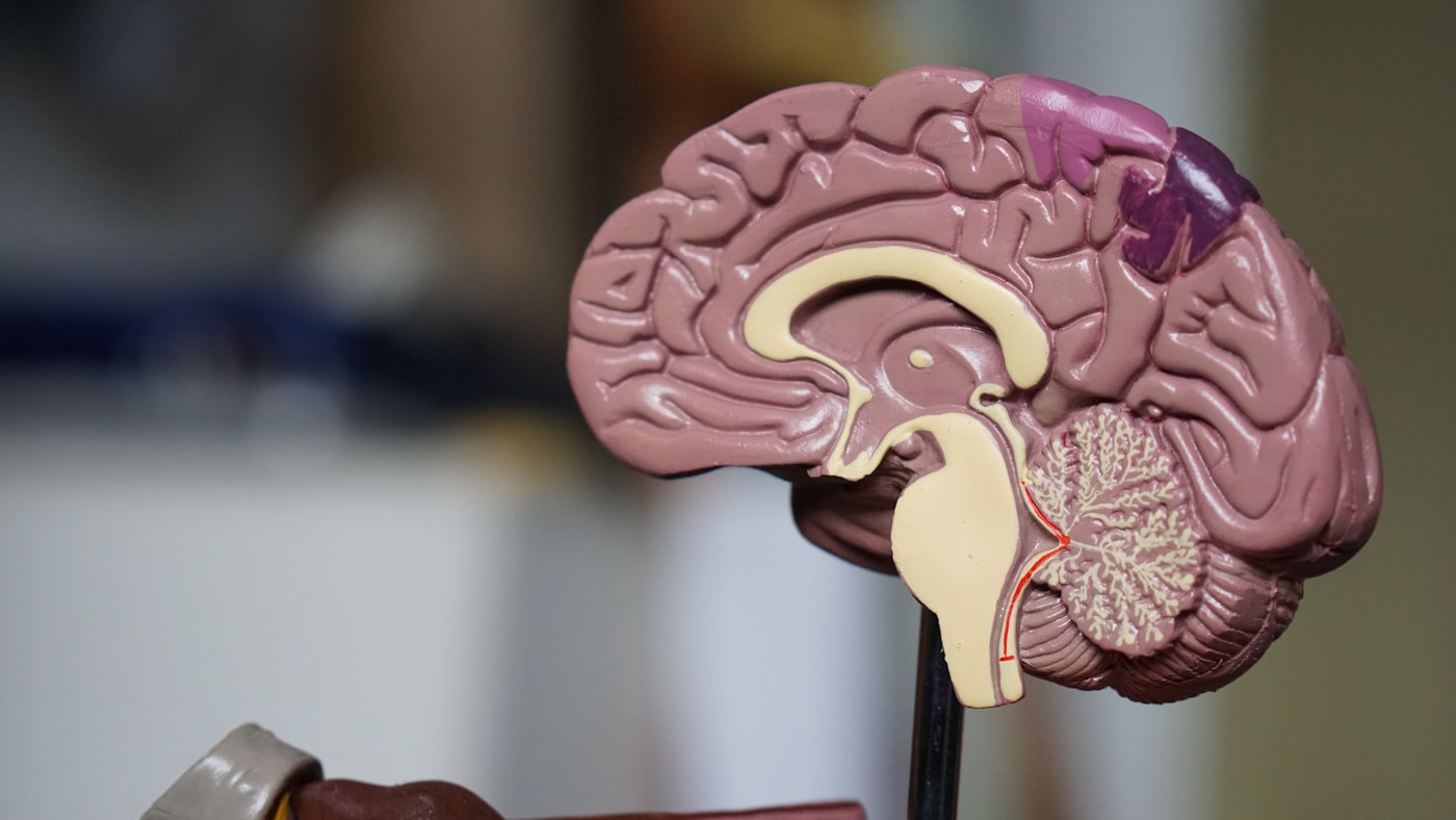
Blood-Based Biomarkers for Early Parkinson’s Detection
The tragedy of Parkinson’s disease has always been its stealth. By the time tremors appear, 50–80% of dopaminergic neurons in the substantia nigra have already been lost. The disease has been burning for a decade or more while remaining clinically invisible.
But we’re entering a new era. Advances in liquid biopsy technology and our understanding of peripheral-to-brain communication — including the groundbreaking STING-mediated extracellular vesicle pathways identified by Härtlova and colleagues at the University of Gothenburg — are revealing that the blood carries molecular whispers of neurodegeneration years before symptoms manifest.
Your blood is no longer just a carrier of oxygen and nutrients. It’s a diagnostic oracle.
The Alpha-Synuclein Revolution
The protein alpha-synuclein has long been Parkinson’s molecular signature — its misfolded aggregates form the Lewy bodies that define the disease pathologically. The challenge was detecting these aggregates reliably in blood rather than cerebrospinal fluid.
That changed with seed amplification assays (SAA). Research from the Michael J. Fox Foundation’s landmark PPMI (Parkinson’s Progression Markers Initiative) study demonstrated that alpha-synuclein SAA can identify Parkinson’s patients with 87.7% sensitivity and 96.3% specificity from blood samples alone.
Even more remarkable: the assay detected pathology in individuals carrying genetic risk variants — LRRK2 and GBA mutations — before any motor symptoms appeared.
- Andrew Siderowf, MD at the University of Pennsylvania led key validation studies showing SAA positivity in prodromal patients
- The test works by amplifying tiny amounts of misfolded protein until they’re detectable — essentially giving pathological seeds room to grow in vitro
- Commercial availability is expanding through companies like Amprion and the Banner Sun Health Research Institute
💡 Quick Fact: In PPMI data published in The Lancet Neurology (2023), alpha-synuclein SAA detected 86% of individuals who would develop Parkinson’s up to 7 years before diagnosis — a detection window that could transform preventive intervention.
What This Means For You
If you carry known genetic risk factors for Parkinson’s (family history of the disease, Ashkenazi Jewish ancestry with higher GBA variant prevalence, or confirmed LRRK2 status), alpha-synuclein seed amplification testing is moving from research into clinical availability. Discuss prodromal screening with a movement disorder specialist — early detection opens windows for neuroprotective lifestyle interventions and emerging disease-modifying trials.
Extracellular Vesicles: The New Frontier
The 2026 research from Öberg, Härtlova, and their team at the Sahlgrenska Academy has added a critical new dimension to blood-based Parkinson’s detection. Their work reveals that STING-dependent peripheral inflammaging drives neurodegeneration via extracellular vesicles — essentially, inflammatory signals from the body’s periphery hijack these tiny membrane-bound parcels to deliver neurotoxic cargo directly to the brain.
This finding transforms our understanding of what blood biomarkers can reveal. Extracellular vesicles (EVs) aren’t just passive debris — they’re active messengers carrying:
- Misfolded proteins including alpha-synuclein oligomers
- Inflammatory cytokines and chemokines that prime microglia for neurotoxic activation
- microRNAs that can reprogram gene expression in recipient neurons
- Mitochondrial fragments signaling peripheral metabolic dysfunction
The Gothenburg team demonstrated that peripheral inflammatory states — the “inflammaging” we’ve discussed throughout this guide — generate EVs that cross the blood-brain barrier and accelerate neurodegeneration. This means systemic inflammatory biomarkers may predict brain-specific disease years in advance.
Crescitelli and colleagues at the Wallenberg Centre for Molecular and Translational Medicine contributed proteomic analysis showing distinct EV signatures in early neurodegeneration — signatures detectable through standard blood draws.
The Inflammatory Panel Approach
Beyond protein aggregation, chronic inflammation itself predicts Parkinson’s risk. Research from Alberto Bhay’s group at Cambridge and the Karolinska Institute’s longitudinal cohorts has identified a constellation of inflammatory markers that elevate years before diagnosis:
- High-sensitivity C-reactive protein (hs-CRP): Elevations above 2.0 mg/L associated with 30% increased Parkinson’s risk over 15 years
- Interleukin-6 (IL-6): Persistent elevation correlates with faster prodromal progression
- Neurofilament light chain (NfL): A marker of axonal damage now measurable in blood, elevated in prodromal Parkinson’s and predictive of conversion from REM sleep behavior disorder
- GDF-15: A mitochondrial stress marker increasingly linked to neurodegeneration risk
The STING pathway research adds interferon-stimulated gene signatures to this panel. When peripheral immune cells chronically activate STING in response to cytoplasmic DNA (from damaged mitochondria, cellular stress, or low-grade infections), they produce a distinctive interferon signature — detectable through gene expression profiling — that correlates with accelerated brain aging.
What This Means For You
Request a comprehensive inflammatory panel as part of your annual longevity assessment. Beyond standard hs-CRP, emerging panels from specialty laboratories now include NfL, IL-6, and GDF-15. While not yet diagnostic for Parkinson’s specifically, persistent inflammatory elevation — especially combined with sleep disturbances or subtle olfactory changes — warrants deeper investigation and aggressive anti-inflammatory lifestyle intervention.
Practical Blood Biomarker Protocol
For those serious about early detection, consider a tiered approach:
Tier 1 — Annual baseline (everyone over 45):
- hs-CRP, fasting insulin, HbA1c
- Complete metabolic panel with lipids
- Homocysteine and vitamin B12
Tier 2 — Enhanced inflammatory assessment (those with risk factors):
- IL-6, TNF-alpha, GDF-15
- Neurofilament light chain (NfL)
- Specialized EV analysis where available
Tier 3 — Prodromal screening (genetic risk or early warning signs):
- Alpha-synuclein seed amplification assay
- STING pathway gene expression profiling
- Serial monitoring every 6–12 months
💡 Quick Fact: REM sleep behavior disorder — acting out dreams physically — converts to Parkinson’s or related synucleinopathies in over 80% of cases within 15 years. If you or your partner notices this, blood-based biomarker screening becomes especially valuable.
Key Points
- Alpha-synuclein seed amplification assays can detect Parkinson’s pathology 5–7 years before symptoms with high sensitivity and specificity — transforming early detection from aspiration to clinical reality
- Extracellular vesicles carry inflammatory and neurotoxic cargo from periphery to brain, as demonstrated by Härtlova’s STING pathway research; EV profiling represents the next frontier in blood-based neurodegeneration screening
- A tiered biomarker approach combining inflammatory markers, NfL, and emerging EV/SAA testing allows personalized risk stratification — enabling intervention during the critical window when neuroprotection remains possible
Emerging Therapies Targeting the Peripheral Inflammation Pathway

Emerging Therapies Targeting the Peripheral Inflammation Pathway
The recognition that peripheral inflammation drives neurodegeneration — rather than simply accompanying it — has catalyzed a paradigm shift in drug development. We’re no longer just trying to protect neurons after damage begins. We’re intercepting the inflammatory cascade at its source, often years before symptoms emerge.
This represents one of the most promising therapeutic frontiers in longevity medicine. And the targets are surprisingly accessible.
STING Inhibitors: Silencing the Inflammatory Alarm
The STING pathway has emerged as a master switch for age-related inflammation. When activated inappropriately — by mitochondrial DNA leakage, cellular stress, or accumulating damage — it triggers the inflammatory cascade that Härtlova’s team at the University of Gothenburg demonstrated travels to the brain via extracellular vesicles.
Several STING inhibitors are now in development:
- H-151 — a potent covalent inhibitor showing efficacy in preclinical neurodegeneration models
- SN-011 — developed by researchers at UT Southwestern, demonstrating blood-brain barrier penetration
- Compound 18 — from GlaxoSmithKline’s pipeline, currently in Phase I trials for inflammatory conditions
The challenge lies in calibrating these interventions. STING plays legitimate roles in immune defense against pathogens and cancer surveillance. Complete suppression isn’t the goal — restoring youthful signaling dynamics is.
💡 Quick Fact: By age 70, baseline STING pathway activation increases by approximately 300% compared to age 30 — even in the absence of infection. This “sterile inflammation” represents a primary driver of inflammaging.
What This Means For You
STING-targeted therapies remain investigational, but understanding this pathway empowers immediate action. Compounds that naturally modulate STING signaling — including omega-3 fatty acids, sulforaphane, and NAD+ precursors — may offer partial benefits while pharmaceutical options mature.
Extracellular Vesicle Interception Strategies
If EVs serve as the delivery vehicles carrying inflammatory cargo to the brain, disrupting this transport represents an elegant therapeutic approach. Researchers are pursuing multiple angles.
Current EV-targeting strategies include:
- Uptake inhibitors — blocking the receptors neurons use to internalize toxic EVs
- Cargo modification — altering the loading process so EVs carry protective rather than destructive payloads
- Clearance enhancement — accelerating hepatic and splenic filtration of circulating EVs
- Designer EVs — engineered vesicles carrying anti-inflammatory or neurotrophic factors
Dr. Miryam Kurzawa-Akanbi’s work at Newcastle University has shown that EV composition shifts dramatically with age and disease. This opens the possibility of EV-based therapeutics — essentially replacing aged, inflammatory vesicles with young, protective ones.
The parallels to young blood plasma research are striking. But EVs offer more precision, carrying specific molecular instructions rather than the complex soup of thousands of plasma proteins.
Senolytic and Senostatic Approaches
Senescent cells — the inflammatory zombies that accumulate with age — are prolific producers of toxic EVs and STING-activating signals. Eliminating or silencing them attacks peripheral inflammaging at its root.
The senolytic landscape has expanded rapidly:
- Dasatinib + Quercetin (D+Q) — the original combination, now in multiple Phase II trials including Unity Biotechnology’s UBX1325 for retinal disease
- Fisetin — a naturally occurring flavonoid showing promise in the AFFIRM-LITE trial at Mayo Clinic under Dr. James Kirkland
- Navitoclax derivatives — more targeted Bcl-2 family inhibitors reducing off-target platelet effects
- CAR-T senolytic approaches — engineered immune cells specifically targeting senescent cell surface markers, pioneered at Memorial Sloan Kettering
Recent data from the Translational Geroscience Network suggests that even intermittent senolytic treatment — monthly or quarterly dosing — substantially reduces circulating inflammatory markers and EV-associated cytokines.
What This Means For You
While prescription senolytics await approval, fisetin (100mg daily or periodic higher doses) and quercetin (500–1000mg) are available now. Combined with time-restricted eating — which naturally promotes senescent cell clearance through autophagy — these represent actionable interventions targeting the peripheral inflammation pathway.
Key Points
- STING inhibitors represent a new therapeutic class targeting the master inflammatory switch that drives EV-mediated neurodegeneration; multiple candidates are advancing through clinical trials
- Extracellular vesicle interception strategies — including uptake blockers, cargo modifiers, and designer replacement EVs — offer precise intervention points in the peripheral-to-brain inflammation highway
- Senolytic therapies eliminate the cellular sources of inflammatory EVs and STING activation; fisetin and quercetin provide accessible options while more potent pharmaceuticals complete development
✦ McKaizer Institute Protocol
Evidence-ranked, actionable steps distilled from the research above.
- Step 1: See the detailed protocol section above.
- Step 2: See the detailed protocol section above.
- Step 3: See the detailed protocol section above.
- Step 4: See the detailed protocol section above.
- Step 5: See the detailed protocol section above.






- Peripheral Inflammation and Parkinson’s Disease How Extracellular Vesicles…
- How Chronic Inflammation Accelerates Aging and What Science Reveals About Reversing It
- How Chronic Inflammation Accelerates Aging and What New Research Reveals About…
- Montana’s Right to Try Law Opens New Frontiers for Experimental Longevity Therapies
- How Stem Cell Exosomes Could Revolutionize Liver Disease Treatment and Longevity





Leave A Comment