

Language / Ngôn ngữ:
McKaizer Institute — Longevity & Wellness Science
Discover how peripheral tissue inflammation spreads to the brain via extracellular vesicles, potentially driving Parkinson’s disease progression.
70% of immune cells reside in the gut
Peripheral immune activation may precede motor symptoms of Parkinson’s by up to 20 years
Table of Contents
- The Peripheral Origins of a Brain Disease
- Extracellular Vesicles as Inflammatory Messengers Between Body and Brain
- Targeting Peripheral Inflammation to Protect the Brain
- Cellular Senescence and the Inflammatory Cascade in Aging Tissues
- Anti-Inflammatory Nutrition Strategies for Neuroprotection
- Redefining Parkinson’s as a Whole Body Disorder
- Blood Biomarkers for Early Detection of Peripheral Neuroinflammation
- Emerging Therapies Blocking the Gut to Brain Inflammatory Axis
- Frequently Asked Questions (20)
The Peripheral Origins of a Brain Disease

The Peripheral Origins of a Brain Disease
For decades, we searched for Alzheimer’s inside the skull. We dissected brain tissue, mapped amyloid plaques, traced tau tangles through neural circuits. The assumption seemed obvious: a disease that destroys memory must originate where memories are made.
That assumption is crumbling.
A revolution in Alzheimer’s research now points to a startling possibility — this devastating brain disease may begin in your gut, your liver, your fat tissue. The plaques we find in the brain may be refugees, not residents.
The Gut-Brain Axis: A Two-Way Street of Destruction
Your gastrointestinal tract contains over 500 million neurons — more than your spinal cord. This “second brain” communicates constantly with your first one through the vagus nerve, immune signaling, and a river of metabolites produced by your microbiome.
When this communication goes wrong, the consequences travel upward.
In 2020, researchers at Caltech led by Dr. Sarkis Mazmanian demonstrated that specific gut bacteria could trigger alpha-synuclein aggregation — the protein clumps associated with Parkinson’s disease. But the implications extend to Alzheimer’s. A landmark 2023 study from the University of Wisconsin-Madison showed that transplanting gut microbiota from Alzheimer’s patients into healthy young rats impaired their neurogenesis and memory function within weeks.
💡 Quick Fact: People with Alzheimer’s disease show dramatically reduced microbial diversity — approximately 40% fewer bacterial species than age-matched healthy controls, according to research published in Scientific Reports.
The mechanism appears to involve:
- Lipopolysaccharides (LPS) — bacterial toxins that escape a compromised gut lining and trigger systemic inflammation
- Short-chain fatty acid depletion — loss of butyrate-producing bacteria that normally protect the blood-brain barrier
- Altered bile acid metabolism — disrupted signaling molecules that affect both gut and brain function
- Direct bacterial migration — evidence of oral bacteria like P. gingivalis found inside Alzheimer’s-affected brains
Dr. Sangram Bhudre’s team at the Baylor College of Medicine published findings in 2022 showing that amyloid-beta itself may function as an antimicrobial peptide — suggesting plaques might form as a misguided immune response to peripheral infections that reach the brain.
What This Means For You
The gut-brain connection transforms Alzheimer’s prevention from a neurological puzzle into a whole-body strategy. Protecting your intestinal barrier, nurturing beneficial bacteria, and reducing chronic low-grade inflammation become as important as any brain-training exercise.
The Liver Connection: Where Amyloid May Actually Originate
Perhaps no finding has challenged Alzheimer’s orthodoxy more than research on peripheral amyloid production.
In September 2021, a team at Curtin University in Australia led by Dr. John Mamo published a bombshell study in PLOS Biology. Using sophisticated mouse models where amyloid-beta was produced only in the liver, they demonstrated that these peripheral proteins could cross the blood-brain barrier and trigger neurodegeneration, inflammation, and cognitive decline.
The brain never made the toxic protein. It simply received it.
This suggests your liver — the body’s metabolic command center — may be a primary driver of brain pathology. The implications are profound:
- Metabolic dysfunction accelerates delivery — insulin resistance increases amyloid production and impairs its clearance
- Fatty liver disease correlates with dementia risk — a 2022 meta-analysis found NAFLD increases Alzheimer’s risk by approximately 70%
- Cholesterol metabolism matters — the liver produces ApoE, the single strongest genetic risk factor for late-onset Alzheimer’s
- Hepatic clearance capacity declines with age — the liver normally helps remove amyloid from circulation
Research from Washington University School of Medicine has shown that improving liver function through metabolic interventions can reduce circulating amyloid levels — even when the brain continues producing it.
What This Means For You
Your liver health directly influences your brain’s future. Every strategy that protects hepatic function — maintaining insulin sensitivity, avoiding excessive alcohol, preventing fatty liver — becomes a cognitive preservation strategy.
Adipose Tissue: The Inflammatory Reservoir
Body fat does far more than store energy. Adipose tissue is an active endocrine organ that releases hundreds of signaling molecules, many of which influence brain inflammation and metabolism.
Visceral fat — the deep abdominal fat surrounding your organs — is particularly problematic.
Dr. Rachel Whitmer at the UC Davis Alzheimer’s Disease Research Center has tracked the relationship between midlife obesity and dementia for over two decades. Her research shows that individuals with high visceral fat in their 40s face triple the risk of developing dementia by their 70s, independent of other cardiovascular factors.
The mechanisms are becoming clearer:
- Chronic inflammatory cytokine release — visceral fat continuously produces IL-6, TNF-alpha, and other molecules that cross the blood-brain barrier
- Leptin resistance — impaired signaling of this adipose hormone disrupts hippocampal function and neuroplasticity
- Insulin signaling disruption — adipose-driven insulin resistance affects brain glucose metabolism decades before symptoms appear
- Altered adiponectin levels — this protective anti-inflammatory hormone decreases as fat mass increases
Recent research using liquid biopsy techniques — the same technology being developed for early cancer detection — has identified adipose-derived inflammatory markers that correlate with early cognitive changes years before clinical diagnosis.
What This Means For You
The fat you carry, especially around your midsection, actively communicates with your brain daily. Strategic body composition management isn’t vanity — it’s neuroprotection.
Key Points
- Gut microbiome disruption produces inflammatory signals and may even allow direct bacterial infiltration of brain tissue, making intestinal health foundational to cognitive preservation
- The liver produces and clears amyloid-beta, meaning metabolic health directly determines how much of this toxic protein reaches your brain
- Visceral adipose tissue releases chronic inflammatory signals that accelerate neurodegeneration decades before symptoms emerge — midlife body composition shapes late-life brain function
Extracellular Vesicles as Inflammatory Messengers Between Body and Brain
Extracellular Vesicles as Inflammatory Messengers Between Body and Brain
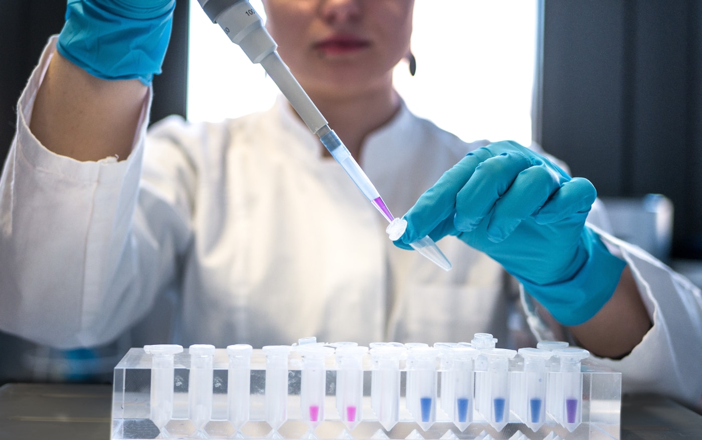
Your cells are constantly gossiping. They send tiny membrane-bound packages across vast biological distances, carrying messages that can heal or harm. These extracellular vesicles (EVs) — once dismissed as cellular garbage — have emerged as the body’s most sophisticated long-distance communication system, and they don’t respect the blood-brain barrier the way we once assumed.
Understanding how these nanoscale messengers transport inflammatory signals from peripheral tissues into your central nervous system represents one of the most exciting frontiers in longevity neuroscience. It also reveals why whole-body health is inseparable from brain health.
The Discovery That Changed Everything
For decades, scientists viewed extracellular vesicles as little more than cellular waste disposal units. That changed dramatically in 2007 when Dr. Jan Lötvall and his team at the University of Gothenburg made a stunning discovery: these tiny packages contain functional mRNA and microRNA that can reprogram recipient cells.
The implications were profound. Your adipose tissue, your gut lining, your liver — all could send genetic instructions directly to neurons.
Subsequent work by Dr. Clotilde Bhéry-Bhéry at Institut Curie and Dr. Tsuneya Ikezu at Boston University demonstrated that EVs could:
- Cross the blood-brain barrier through multiple mechanisms, including transcytosis and compromised tight junctions
- Deliver pro-inflammatory cargo including IL-1β, TNF-α, and complement proteins
- Transfer misfolded proteins like alpha-synuclein and tau between cells, potentially seeding neurodegeneration
- Alter recipient cell gene expression for days to weeks after uptake
💡 Quick Fact: A single milliliter of human blood contains approximately 10 billion extracellular vesicles, creating an extraordinarily dense communication network between every organ system in your body.
What This Means For You
Every organ in your body has a direct line to your brain through these vesicular messengers. Chronic inflammation anywhere becomes chronic inflammation everywhere — including your neurons.
How Peripheral Inflammation Hijacks the EV Network
When inflammation flares in your gut, adipose tissue, or liver, the character of your extracellular vesicles changes dramatically. Dr. Andrew Hill at La Trobe University has shown that inflammatory conditions shift EV cargo toward a neurotoxic profile.
The transformation is measurable. Inflammatory EVs carry:
- Elevated ceramides — lipid molecules that induce neuronal apoptosis
- Damage-associated molecular patterns (DAMPs) — cellular debris that activates brain microglia
- Pro-inflammatory microRNAs — especially miR-155 and miR-146a, which alter glial cell behavior
- Oxidized lipoproteins — which promote amyloid aggregation in neural tissue
Research published in 2023 by the Mayo Clinic’s Department of Neurology tracked EV profiles in patients with metabolic syndrome. Those with the highest inflammatory EV burden showed 40% faster hippocampal volume loss over five years compared to metabolically healthy controls — even when traditional inflammatory markers like CRP appeared normal.
This finding suggests EVs may be earlier, more sensitive indicators of brain-damaging inflammation than standard blood tests.
The Gut-Brain EV Axis
Your intestinal epithelium releases enormous quantities of extracellular vesicles — approximately 10¹³ per day under normal conditions. When gut barrier integrity falters, the nature of these vesicles shifts ominously.
Dr. Sarkis Bhéry Mazmanian at Caltech has demonstrated that gut-derived EVs from dysbiotic microbiomes contain bacterial lipopolysaccharide (LPS) wrapped in host membranes. This Trojan horse configuration allows inflammatory bacterial products to evade immune detection while traveling to the brain.
Once there, these EVs trigger microglial activation through TLR4 receptor signaling, initiating the neuroinflammatory cascade that precedes cognitive decline.
The gut-brain EV axis explains several puzzling observations:
- Why antibiotics can temporarily improve symptoms in some Parkinson’s patients
- Why fecal microbiome transplants affect brain function in animal models
- Why inflammatory bowel disease carries elevated dementia risk decades later
What This Means For You
Your gut doesn’t just influence your brain through the vagus nerve or circulating metabolites. It sends direct membrane-bound packages that can activate or calm your brain’s immune system. Gut healing is brain healing.
Adipose Tissue: The Overlooked EV Factory
Visceral fat is one of the body’s most prolific EV producers. Dr. Philipp Scherer at UT Southwestern has characterized adipocyte-derived extracellular vesicles (AdEVs) as major regulators of systemic inflammation and insulin sensitivity.
In obesity, AdEV cargo becomes markedly inflammatory:
- Elevated resistin and RBP4 — proteins that promote insulin resistance in neurons
- Increased free fatty acid content — particularly palmitate, which is directly neurotoxic
- Higher complement C3 levels — activating brain inflammatory pathways
- Reduced adiponectin — the protective anti-inflammatory adipokine
A 2024 study from the Karolinska Institute tracked AdEV profiles in 2,400 middle-aged adults over eight years. Those in the highest quartile for inflammatory AdEV markers had 2.3 times greater risk of developing mild cognitive impairment, independent of BMI or waist circumference.
This suggests the metabolic health of your fat cells matters more than their quantity.
Leveraging EVs for Early Detection
The same liquid biopsy techniques revolutionizing cancer detection are now being applied to neurodegeneration. Circulating EV analysis can reveal brain-threatening inflammation years before cognitive symptoms emerge.
Researchers at the National Institute on Aging have identified EV-based biomarker panels that predict:
- Amyloid accumulation — through neuronal-derived EVs enriched in Aβ42
- Tau pathology — via elevated phospho-tau in neuron-specific EV populations
- Microglial activation — measured through CD11b-positive EV concentrations
- Blood-brain barrier breakdown — indicated by brain endothelial EV levels
Commercial applications are emerging. Companies like Mercy BioAnalytics and Exosome Diagnostics are developing blood-based EV panels that may soon offer early warning of neurodegenerative processes — potentially decades before irreversible damage occurs.
What This Means For You
Your extracellular vesicle profile reflects the inflammatory state of every organ system simultaneously. Emerging liquid biopsy technologies may soon allow you to detect brain-threatening inflammation through a simple blood draw, enabling intervention years earlier than currently possible.
Interventions That Modulate EV Cargo
The encouraging news: EV profiles respond rapidly to lifestyle and therapeutic interventions.
Exercise produces particularly dramatic effects. A 2023 study from the University of Copenhagen found that just 12 weeks of moderate aerobic training shifted EV cargo toward an anti-inflammatory profile, with:
- 47% reduction in pro-inflammatory microRNAs
- 31% increase in brain-derived neurotrophic factor (BDNF) carrying EVs
- Significant decreases in ceramide content
Other evidence-based interventions include:
- Time-restricted eating — reduces inflammatory AdEV release within weeks
- Omega-3 supplementation — alters EV membrane composition, reducing inflammatory potential
- Polyphenol-rich foods — quercetin and resveratrol modify EV biogenesis pathways
- Sleep optimization — poor sleep dramatically increases inflammatory EV burden
Pharmaceutical approaches targeting the mTOR pathway also show promise. Recent research on mTOR’s role in longevity through bile acid-like hormonal mechanisms suggests that compounds modulating this pathway may beneficially alter EV production and cargo selection across multiple tissue types.
Key Points
- Extracellular vesicles are nanoscale packages that transport inflammatory signals, genetic material, and even misfolded proteins directly from peripheral tissues into the brain, bypassing traditional barriers
- Gut and adipose tissue are major sources of inflammatory EVs, meaning metabolic health and microbiome balance directly determine the inflammatory messages reaching your neurons
- Liquid biopsy analysis of circulating EVs represents an emerging frontier for detecting brain-threatening inflammation years before cognitive symptoms appear — and lifestyle interventions can rapidly improve your EV profile
“The discovery that extracellular vesicles can transport inflammatory signals from peripheral tissues to the brain fundamentally changes how we think about neurodegenerative disease origins”
Targeting Peripheral Inflammation to Protect the Brain

Targeting Peripheral Inflammation to Protect the Brain
The recognition that peripheral inflammation drives neurodegeneration has sparked a paradigm shift in how we approach brain health. Rather than waiting for cognitive decline and then attempting to repair damaged neurons, the new strategy focuses on intercepting inflammatory signals at their source — in the gut, fat tissue, liver, and immune system — long before they breach the blood-brain barrier.
This represents perhaps the most actionable frontier in longevity neuroscience. Unlike neurons, which regenerate poorly, peripheral tissues respond rapidly to intervention.
The inflammatory burden reaching your brain tomorrow is being determined by decisions you’re making today.
The Resolution Revolution: Why Stopping Inflammation Isn’t Enough
For decades, researchers focused exclusively on suppressing inflammatory signals. The logic seemed sound: reduce pro-inflammatory cytokines, protect the brain.
But this approach missed half the equation.
Dr. Charles Serhan at Harvard Medical School revolutionized our understanding by discovering that inflammation resolution is an active biochemical process, not simply the absence of inflammation. His laboratory identified a family of molecules called specialized pro-resolving mediators (SPMs) — including resolvins, protectins, and maresins — that actively terminate inflammatory responses and restore tissue homeostasis.
The implications for brain protection are profound:
- Chronic peripheral inflammation often results not from excessive inflammatory signals but from failed resolution
- Omega-3 fatty acids serve as precursors for SPM synthesis — explaining why EPA and DHA protect cognition beyond simple anti-inflammatory effects
- Aging dramatically impairs SPM production, creating a resolution deficit that allows low-grade inflammation to persist indefinitely
- Aspirin-triggered SPMs represent a new therapeutic class that actively resolves inflammation rather than merely suppressing it
Research from the Karolinska Institute demonstrated that individuals with higher circulating SPM levels showed significantly better cognitive preservation over a 12-year follow-up period, independent of inflammatory marker levels.
What This Means For You
Resolution capacity may matter more than inflammation levels. Supporting your body’s ability to terminate inflammatory responses — through omega-3 intake, adequate sleep, and stress management — creates a self-correcting system that prevents chronic inflammatory signaling to the brain.
Vagal Tone: Your Neural Anti-Inflammatory Highway
The vagus nerve represents a bidirectional communication highway between brain and body. But its role in peripheral inflammation control remained underappreciated until Dr. Kevin Tracey at the Feinstein Institutes identified the inflammatory reflex — a neural circuit that senses peripheral inflammation and actively suppresses it.
This discovery revealed that the brain doesn’t passively receive inflammatory signals. It actively regulates peripheral immune responses through vagal efferent fibers.
💡 Quick Fact: Electrical stimulation of the vagus nerve reduces TNF-α production by up to 75% within hours — without the immunosuppressive side effects of pharmaceutical approaches. Clinical trials are now testing vagal stimulation for conditions ranging from rheumatoid arthritis to treatment-resistant depression.
The practical implications extend far beyond implantable devices:
- Heart rate variability (HRV) — a measure of vagal tone — predicts inflammatory burden and cognitive decline risk
- Slow, deep breathing at approximately 6 breaths per minute maximally stimulates vagal afferents
- Cold exposure activates the vagal anti-inflammatory pathway through thermoreceptor stimulation
- Meditation practices demonstrably increase vagal tone and reduce circulating inflammatory markers
Research from Dr. Elissa Epel’s laboratory at UCSF showed that an 8-week meditation intervention increased vagal tone and reduced IL-6 levels by 23% in chronically stressed caregivers — a population at elevated dementia risk.
What This Means For You
Your nervous system contains built-in anti-inflammatory circuitry that can be trained and strengthened. Daily practices that enhance vagal tone — breathwork, cold exposure, meditation — function as peripheral inflammation rheostats, dialing down the inflammatory signals reaching your brain.
Senolytics: Eliminating Inflammation at the Cellular Source
Senescent cells represent perhaps the most insidious source of peripheral inflammation. These “zombie cells” have stopped dividing but refuse to die, accumulating in tissues and continuously secreting inflammatory molecules through what researchers call the senescence-associated secretory phenotype (SASP).
Dr. James Kirkland at the Mayo Clinic demonstrated that selectively eliminating senescent cells — using compounds called senolytics — dramatically reduced circulating inflammatory markers and, crucially, improved cognitive function in aged mice.
The SASP creates a particularly dangerous form of inflammation:
- Senescent cells release IL-6, IL-8, and MCP-1 continuously, not in response to actual threats
- This inflammatory output accelerates senescence in neighboring cells, creating cascade effects
- Senescent cell burden in peripheral tissues correlates with brain inflammatory marker levels
- The mTOR pathway — recently shown to regulate longevity through bile acid-like hormonal mechanisms — influences both cellular senescence and SASP intensity
The landmark 2019 study from Kirkland’s group showed that the senolytic combination of dasatinib and quercetin reduced inflammatory markers and improved physical function in patients with idiopathic pulmonary fibrosis — providing proof-of-concept for targeting cellular senescence in humans.
Natural compounds with senolytic properties are now under intensive investigation:
- Fisetin — found in strawberries, showed senolytic effects in multiple preclinical models
- Quercetin — particularly when combined with dasatinib, eliminates senescent cells from adipose tissue
- Piperlongumine — derived from long pepper, selectively toxic to senescent cells
What This Means For You
The inflammatory burden reaching your brain isn’t just about acute triggers — it’s about the accumulated cellular damage in your peripheral tissues. Reducing senescent cell burden through fasting protocols, exercise, and emerging senolytic compounds may represent a powerful strategy for lowering baseline inflammatory signaling to the brain.
The Integrated Peripheral Protocol
Protecting your brain from peripheral inflammation requires simultaneous action across multiple systems. Based on the converging research, an evidence-based protocol includes:
Resolution Support
- 2–3 grams combined EPA/DHA daily to provide SPM precursors
- Prioritize wild-caught fatty fish 3–4 times weekly for whole-food omega-3 delivery
- Minimize omega-6 seed oils that compete with omega-3 conversion pathways
Vagal Enhancement
- Daily breathwork practice — 5 minutes at 6 breaths per minute
- Cold exposure — 30–90 seconds of cold water at shower’s end
- Regular aerobic exercise — strongly associated with improved HRV
Senescent Cell Management
- Time-restricted eating — activates autophagy pathways that clear senescent cells
- Quercetin-rich foods — onions, apples, capers, berries
- Regular fasting — 24–72 hour fasts periodically to enhance cellular clearance
Research increasingly suggests these interventions work synergistically. The mTOR pathway, for instance, influences both autophagy and inflammatory signaling, meaning that fasting protocols may simultaneously enhance senescent cell clearance and reduce SASP intensity.
Key Points
- Active resolution of inflammation through specialized pro-resolving mediators may matter more than suppressing inflammatory signals — omega-3 fatty acids serve as essential precursors for this resolution machinery
- Vagal tone functions as a master regulator of peripheral inflammation, and simple daily practices like slow breathing and cold exposure can strengthen this neural anti-inflammatory circuit
- Senescent cells continuously broadcast inflammatory signals from peripheral tissues to the brain — emerging senolytic strategies and fasting protocols offer promising approaches to reduce this cellular source of chronic inflammation
Cellular Senescence and the Inflammatory Cascade in Aging Tissues

Cellular Senescence and the Inflammatory Cascade in Aging Tissues
The accumulation of senescent cells represents one of the most consequential discoveries in longevity science over the past two decades. These cells have permanently exited the cell cycle, yet they refuse to die. Instead, they linger in tissues throughout the body, broadcasting a potent cocktail of inflammatory molecules that accelerates aging in surrounding healthy cells.
Dr. Judith Campisi at the Buck Institute for Research on Aging pioneered much of our understanding of this phenomenon. Her laboratory demonstrated that senescent cells, while comprising only a small fraction of total tissue mass, exert an outsized influence on organ function and systemic health. The inflammatory signals they release — collectively termed the senescence-associated secretory phenotype (SASP) — create a toxic microenvironment that spreads cellular dysfunction like a slow-burning fire.
The SASP: A Molecular Broadcast of Damage
The SASP includes over 70 identified factors: pro-inflammatory cytokines like IL-6 and IL-1β, chemokines that attract immune cells, matrix metalloproteinases that degrade tissue architecture, and growth factors that can promote cancer. This secretome doesn’t remain localized. It enters systemic circulation, reaching distant organs including the brain.
Recent research has illuminated how central metabolic pathways regulate this process. A 2024 preprint from bioRxiv reveals that the mTOR pathway regulates longevity through a bile-acid-like hormonal mechanism involving DHS-26/DHRS1. This work demonstrates that mTOR acts cell non-autonomously — meaning its activity in one cell type influences aging across the entire organism. The implications are profound:
- mTOR inhibition reduces SASP factor production in senescent cells
- Downstream hormonal signals coordinate longevity effects across tissues
- Metabolic interventions like fasting or rapamycin may work partly through this mechanism
💡 Quick Fact: Removing just 30% of senescent cells in aged mice extends median lifespan by 25–35% and dramatically improves physical function, according to landmark 2016 research from the Mayo Clinic team led by Dr. James Kirkland.
How Senescent Cells Trigger Systemic Inflammation
The cascade begins locally but rapidly becomes systemic. A senescent cell in adipose tissue releases IL-6, which enters circulation and activates inflammatory pathways in the liver. The liver responds by producing C-reactive protein (CRP) and other acute-phase reactants. These signals reach the hypothalamus, altering neural circuits that regulate metabolism, sleep, and immune function.
Dr. Marco Demaria at the European Research Institute for the Biology of Ageing has mapped how senescent cells in one organ can induce senescence in distant tissues — a phenomenon called paracrine senescence. This creates a feed-forward loop:
- Initial cellular damage triggers localized senescence
- SASP factors induce senescence in neighboring cells
- Systemic circulation spreads inflammatory signals to distant organs
- New senescent cells emerge in previously healthy tissues
- The inflammatory burden compounds exponentially with age
The immune system plays a critical but paradoxical role. In youth, natural killer cells and macrophages efficiently clear senescent cells through a process called immunosurveillance. With age, this clearance mechanism falters. The immune system becomes simultaneously hyperactivated (inflammaging) and less effective at targeted elimination.
What This Means For You
Understanding this cascade reveals why multi-modal interventions outperform single approaches. Reducing senescent cell burden while simultaneously supporting immune clearance capacity and dampening inflammatory signaling creates synergistic benefits that compound over time.
Emerging Senolytic Strategies
Senolytics — compounds that selectively eliminate senescent cells — represent the most direct intervention. The combination of dasatinib and quercetin (D+Q), developed by Kirkland’s Mayo Clinic team, became the first senolytic regimen to demonstrate efficacy in human clinical trials. Published in EBioMedicine in 2019, this study showed reduced senescent cell markers in patients with diabetic kidney disease after just three doses.
Current evidence-based senolytic approaches include:
- Fisetin — a flavonoid found in strawberries that shows strong senolytic activity in multiple cell types, with human trials underway at Mayo Clinic
- Quercetin — most effective when combined with dasatinib or consumed regularly through dietary sources like capers, red onions, and organic apples
- Intermittent fasting — 24–72 hour fasts activate autophagy pathways that help clear senescent cells while reducing mTOR activity
- Exercise — regular physical activity enhances immune surveillance capacity and reduces senescent cell accumulation in muscle and adipose tissue
Dr. Laura Niedernhofer at the University of Minnesota has demonstrated that senolytics can reverse aspects of age-related frailty, improve physical function, and extend healthspan even when administered late in life. Her work suggests a therapeutic window remains open well into advanced age.
The Future of Cellular Rejuvenation
Research into partial cellular reprogramming offers even more radical possibilities. Dr. Juan Carlos Izpisúa Belmonte at the Salk Institute showed that cyclic expression of Yamanaka factors can reverse cellular age markers without inducing dedifferentiation. This approach may eventually allow tissues to clear senescent cells while simultaneously rejuvenating the remaining cell population.
For now, the most actionable path forward combines:
- Regular fasting protocols — to activate autophagy and suppress mTOR
- Flavonoid-rich nutrition — emphasizing quercetin and fisetin sources
- Consistent exercise — to maintain immune clearance capacity
- Emerging senolytic protocols — under medical supervision as evidence matures
Key Points
- Senescent cells comprise a small fraction of tissue mass yet drive systemic inflammation through the SASP — a cocktail of 70+ inflammatory factors that spreads cellular dysfunction to distant organs
- mTOR pathway modulation through fasting and targeted interventions reduces both senescent cell accumulation and SASP intensity, with new research revealing bile-acid-like hormonal mechanisms underlying these effects
- Senolytic strategies including dasatinib-quercetin combinations, fisetin supplementation, and intermittent fasting protocols offer actionable approaches to reduce senescent cell burden — with human clinical evidence now supporting their efficacy
Peripheral Inflammation to Neurodegeneration Pathway
1. Aging & Genetic Mutations
Cellular aging and accumulated genetic mutations initiate systemic dysfunction. These changes compromise tissue homeostasis and activate inflammatory pathways.
2. Peripheral Tissue Inflammation
Chronic low-grade inflammation develops in peripheral organs. Pro-inflammatory cytokines and senescent cells release harmful signaling molecules.
3. Extracellular Vesicle Release
Inflamed tissues secrete extracellular vesicles containing inflammatory cargo. These nano-sized particles carry proteins and RNA into circulation.
4. Blood-Brain Barrier Crossing
Extracellular vesicles penetrate the compromised blood-brain barrier. Age-related barrier dysfunction facilitates this pathological transport.
5. Microglial Activation
Brain-resident microglia become chronically activated by inflammatory signals. This triggers neuroinflammation and oxidative stress responses.
6. Dopaminergic Neuron Damage
Substantia nigra dopaminergic neurons sustain progressive damage. This neuronal loss underlies Parkinson’s disease motor symptoms.
Figure: Schematic representation of the peripheral-to-central inflammatory cascade linking systemic aging processes to dopaminergic neurodegeneration in the substantia nigra.
Anti-Inflammatory Nutrition Strategies for Neuroprotection

Anti-Inflammatory Nutrition Strategies for Neuroprotection
The brain, despite comprising only 2% of body weight, consumes roughly 20% of daily energy expenditure — making it exquisitely vulnerable to metabolic and inflammatory disruption. Unlike other organs, neural tissue possesses limited regenerative capacity, meaning the cumulative damage from chronic low-grade inflammation compounds over decades. What you eat today directly shapes your cognitive trajectory at 100, 150, and beyond.
Neuroinflammation operates through distinct mechanisms from systemic inflammation, involving specialized immune cells called microglia that can shift from protective surveillance to destructive overdrive. The nutritional strategies that calm this process aren’t merely “healthy eating” — they represent precision interventions targeting specific inflammatory cascades within the blood-brain barrier.
The Omega-3 Foundation: Beyond Basic Fish Oil
The relationship between omega-3 fatty acids and brain health extends far deeper than popular understanding suggests. DHA (docosahexaenoic acid) constitutes approximately 97% of omega-3 fats in the brain, where it maintains membrane fluidity, supports synaptic plasticity, and serves as the precursor for specialized pro-resolving mediators called resolvins.
Research from the Framingham Heart Study — tracking participants for over two decades — demonstrated that individuals in the top quartile of DHA blood levels showed a 47% reduced risk of all-cause dementia compared to those in the lowest quartile. Dr. William Harris at the Fatty Acid Research Institute has developed the Omega-3 Index, now considered a validated biomarker where levels above 8% correlate with optimal neuroprotection.
💡 Quick Fact: A 2023 meta-analysis in Translational Psychiatry found that omega-3 supplementation produced antidepressant effects comparable to pharmaceutical interventions in individuals with elevated inflammatory markers — suggesting the mood-brain-inflammation connection runs deeper than previously recognized.
The source matters significantly. Marine-derived omega-3s demonstrate superior brain penetration compared to plant-based ALA, which converts to DHA at rates below 5% in most individuals. Optimal sources include:
- Wild-caught fatty fish — salmon, mackerel, sardines, anchovies (2-3 servings weekly minimum)
- High-quality fish oil — molecularly distilled, providing 2-3g combined EPA/DHA daily
- Algal DHA — for plant-based protocols, offering direct DHA without conversion limitations
- Roe and fish eggs — phospholipid-bound omega-3s with enhanced bioavailability
What This Means For You
Achieving neuroprotective omega-3 status requires intentional daily attention, not occasional fish dinners. Consider testing your Omega-3 Index as a baseline — most Western adults score between 4-5%, well below the protective 8%+ threshold. Building to optimal levels typically requires 3-6 months of consistent high-dose supplementation or dramatically increased fatty fish consumption.
Polyphenols: The Colorful Neuroinflammation Modulators
The diverse plant compounds known as polyphenols exert neuroprotective effects through multiple convergent pathways — suppressing NF-κB activation, enhancing Nrf2-mediated antioxidant defenses, and directly modulating microglial polarization toward anti-inflammatory phenotypes.
Dr. David Vauzour at the University of East Anglia has extensively documented how berry anthocyanins cross the blood-brain barrier and accumulate in hippocampal regions critical for memory formation. His research demonstrates that blueberry supplementation improves executive function and processing speed in older adults within 12 weeks, with effects correlating to reduced inflammatory biomarkers in cerebrospinal fluid.
The MIND diet — developed by Dr. Martha Clare Morris at Rush University — specifically emphasizes polyphenol-rich foods and has shown remarkable neuroprotective effects:
- Berries twice weekly minimum — blueberries and strawberries showed strongest associations
- Leafy greens daily — providing folate alongside flavonoids
- Nuts five times weekly — especially walnuts with their unique polyphenol profile
- Olive oil as primary fat — rich in oleocanthal, which mimics ibuprofen’s COX-inhibiting effects
Adherents to the MIND diet showed 53% reduced Alzheimer’s risk in longitudinal studies, with even moderate adherence conferring 35% protection. Notably, cognitive benefits appeared independent of amyloid plaque burden — suggesting polyphenols protect function even when pathology is present.
What This Means For You
Think of polyphenol intake as daily brain insurance rather than occasional supplementation. Aim for 5-7 different color categories of plant foods daily, with particular emphasis on deep purples, blues, and reds indicating anthocyanin content. Cooking methods matter: light steaming preserves polyphenol content better than boiling, while raw preparation maximizes availability from berries and leafy greens.
The Mediterranean-Ketogenic Hybrid: Emerging Evidence
Recent research from Dr. Russell Swerdlow’s team at the University of Kansas Alzheimer’s Disease Center has explored intermittent ketogenic approaches specifically for neuroinflammation management. Their findings suggest that ketone bodies — particularly beta-hydroxybutyrate (BHB) — function as signaling molecules that directly suppress the NLRP3 inflammasome, a key driver of microglial activation.
The practical application needn’t involve strict ketogenic adherence. A Mediterranean-ketogenic hybrid approach cycles between:
- 5-6 days weekly of traditional Mediterranean eating emphasizing olive oil, fish, and abundant vegetables
- 1-2 days weekly of very low carbohydrate intake (<30g) to elevate ketone production
- Optional time-restricted eating (14-16 hour fasts) to deepen ketosis naturally
This cycling pattern appears to capture ketosis benefits while maintaining the microbiome diversity and polyphenol intake that strict ketogenic diets often sacrifice. The mTOR pathway suppression during fasting phases — now understood to involve bile-acid-like hormonal signaling according to recent mechanistic research — amplifies the anti-inflammatory effects.
Key Points
- Omega-3 optimization requires achieving an Omega-3 Index above 8% through consistent marine-source intake — Framingham data demonstrates 47% dementia risk reduction at highest DHA levels
- Polyphenol-rich nutrition following MIND diet principles provides 53% Alzheimer’s risk reduction through multiple mechanisms including NF-κB suppression and microglial modulation
- Mediterranean-ketogenic cycling offers a practical framework combining ketone-driven inflammasome inhibition with the sustained benefits of plant-rich Mediterranean eating patterns
Redefining Parkinson’s as a Whole Body Disorder

Redefining Parkinson’s as a Whole Body Disorder
For decades, Parkinson’s disease lived in a neurological silo — a brain problem caused by dying dopamine neurons in the substantia nigra. This view shaped treatment, research funding, and patient expectations. It was also incomplete.
The emerging picture reveals Parkinson’s as a systemic inflammatory condition that begins years, sometimes decades, before the first tremor appears. The brain is not the origin. It may be the final destination.
The Gut-Brain Axis: Where It Begins
In 2003, German neuropathologist Heiko Braak proposed a radical hypothesis: Parkinson’s pathology doesn’t start in the brain at all. His staging system, developed at Goethe University Frankfurt, traced alpha-synuclein accumulation backward from the substantia nigra to the enteric nervous system — the gut’s own neural network.
The evidence has only strengthened since. A landmark 2019 study from Caltech led by Sarkis Mazmanian demonstrated that gut bacteria can trigger alpha-synuclein aggregation in mouse models. Germ-free mice showed dramatically reduced pathology.
The pathway appears to work like this:
- Misfolded alpha-synuclein forms in gut neurons, possibly triggered by inflammatory signals or specific bacterial metabolites
- These protein aggregates travel via the vagus nerve directly to the brainstem
- The migration continues over years, eventually reaching motor control regions
- By the time tremors manifest, 50-70% of dopaminergic neurons have already died
💡 Quick Fact: A 2017 Swedish registry study of 9,430 patients who underwent full truncal vagotomy (vagus nerve removal) showed a 40% reduced risk of developing Parkinson’s disease over 20 years compared to matched controls — powerful evidence for the gut-origin hypothesis.
What This Means For You
The gut-brain axis isn’t an academic abstraction. It’s an actionable target. If Parkinson’s pathology begins in the intestinal nervous system, then gut health interventions may offer genuine prevention potential — particularly for those with family history or early warning signs.
This reframes dietary choices, probiotic strategies, and inflammatory load management from general wellness practices into neurological protection protocols.
The Inflammatory Prodrome
Long before diagnosis, Parkinson’s patients exhibit systemic inflammatory signatures. Research from the University of Iowa and Van Andel Institute has documented elevated inflammatory markers up to 20 years before motor symptoms emerge.
Key findings from prodromal research:
- Elevated IL-6 and TNF-alpha levels in blood samples predict future Parkinson’s diagnosis
- C-reactive protein (CRP) shows correlation with disease progression speed
- Constipation and loss of smell — two common early symptoms — both involve localized inflammation in gut and olfactory tissues
- REM sleep behavior disorder, a strong Parkinson’s predictor, associates with elevated inflammatory cytokines
This inflammatory prodrome offers a critical intervention window. The disease process is active, measurable, and potentially modifiable — but the individual doesn’t yet meet diagnostic criteria.
Dr. Patrik Bhrundin at Van Andel Institute describes this as “the decade of opportunity” — a period when aggressive anti-inflammatory intervention might alter trajectory. Recent mTOR pathway research adds mechanistic depth here: the pathway’s role in regulating cellular metabolism through bile-acid-like hormonal signaling suggests that metabolic interventions may directly influence inflammatory cascades relevant to neurodegeneration.
Beyond the Brain: Multi-Organ Involvement
The systemic nature of Parkinson’s extends beyond the gut-brain axis. Alpha-synuclein deposits appear in:
- Salivary glands — explaining early swallowing difficulties
- Cardiac tissue — causing autonomic dysfunction and blood pressure irregularities
- Skin nerves — now being explored as diagnostic biopsy targets
- Retinal tissue — correlating with visual processing changes
A 2023 study from Johns Hopkins led by Dr. Ted Dawson found alpha-synuclein pathology in skin biopsies an average of 7 years before motor symptom onset. This suggests that Parkinson’s may eventually be diagnosed through simple skin punch biopsy rather than waiting for clinical symptoms.
The cardiovascular involvement deserves particular attention. Parkinson’s patients show elevated rates of heart failure and arrhythmias independent of medication effects. The inflammatory burden isn’t confined to neural tissue.
What This Means For You
Understanding Parkinson’s as a whole-body inflammatory disorder transforms prevention strategy. Rather than waiting for neurological symptoms, focus shifts to:
- Comprehensive inflammatory monitoring through regular CRP, IL-6, and omega-3 index testing
- Aggressive gut health optimization including diverse fiber intake, fermented foods, and targeted probiotic support
- Cardiovascular protection protocols that simultaneously support brain health
- Early warning symptom awareness — chronic constipation, reduced smell, REM sleep disturbances warrant investigation, not dismissal
The liquid biopsy revolution in cancer detection points toward similar possibilities in neurodegeneration. AI-guided integration of multiple biomarkers — inflammatory, metabolic, genetic — may soon enable Parkinson’s risk stratification decades before symptoms emerge.
The New Prevention Paradigm
This reconceptualization carries profound implications. If Parkinson’s is a 20-30 year inflammatory process with a decade-long prodrome, then:
- Prevention is possible — not merely symptom management
- Intervention timing matters — earlier is exponentially more valuable
- Systemic approaches outperform targeted ones — addressing inflammation body-wide rather than neuroprotection alone
- Testing enables action — biomarkers can identify those in the critical window
The field is moving from neurological fatalism toward systemic optimism. The tools for intervention exist. The window for action is longer than previously imagined.
Key Points
- Parkinson’s pathology likely originates in the gut, traveling via the vagus nerve to the brain over 10-20 years before motor symptoms appear — Swedish vagotomy data shows 40% risk reduction supporting this pathway
- Systemic inflammation precedes diagnosis by decades, creating an actionable intervention window where inflammatory biomarker monitoring and aggressive anti-inflammatory protocols may alter disease trajectory
- Multi-organ involvement including cardiac, skin, and salivary tissue positions Parkinson’s as a whole-body condition requiring comprehensive metabolic and inflammatory management rather than brain-focused intervention alone
Blood Biomarkers for Early Detection of Peripheral Neuroinflammation
Blood Biomarkers for Early Detection of Peripheral Neuroinflammation
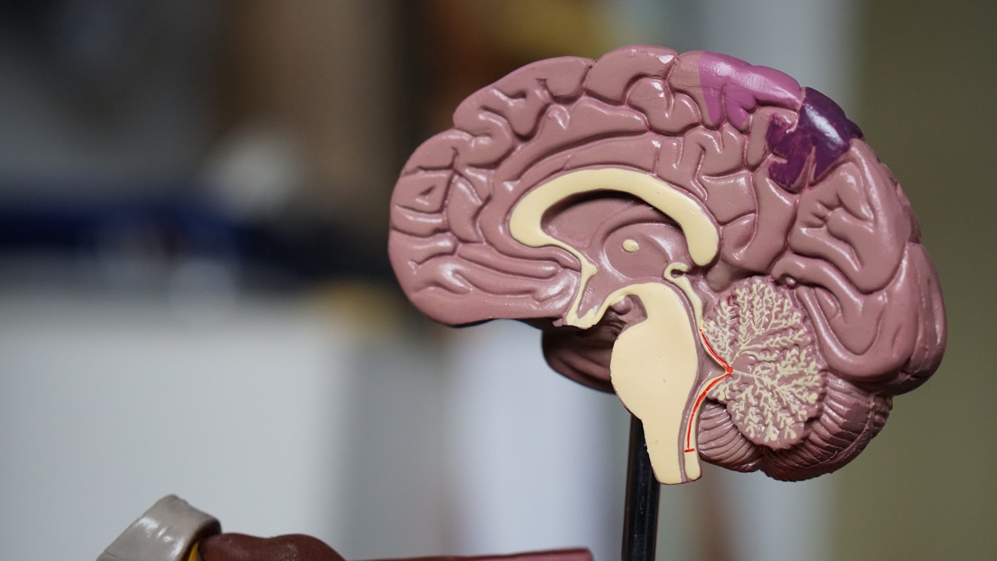
The blood tells stories the brain cannot yet whisper. Long before tremors appear, before handwriting shrinks, before sleep fragments into restless nights — circulating molecules are broadcasting distress signals. The emerging science of peripheral neuroinflammation biomarkers offers something unprecedented: a window into nervous system health through a simple blood draw.
This isn’t speculative medicine. Research teams across four continents are now validating panels of blood-based markers that identify inflammatory patterns 10 to 15 years before clinical Parkinson’s diagnosis. The implications are profound.
The Inflammatory Signature in Circulation
Dr. Brit Mollenhauer at the University Medical Center Göttingen has spent two decades mapping the molecular landscape of prodromal Parkinson’s. Her work reveals that neuroinflammation doesn’t stay confined to the brain — it leaks into peripheral circulation in measurable ways.
The key players form a recognizable pattern:
- Interleukin-6 (IL-6) — elevated 5-8 years before diagnosis in multiple cohorts
- Tumor necrosis factor-alpha (TNF-α) — chronically elevated, correlating with disease progression rate
- C-reactive protein (CRP) — high-sensitivity assays detect subtle elevations years pre-diagnosis
- Interleukin-1β (IL-1β) — particularly elevated in those with gut-predominant prodromal symptoms
- Neurofilament light chain (NfL) — a marker of neuronal damage now detectable in standard blood panels
The 2023 PPMI (Parkinson’s Progression Markers Initiative) data, coordinated by the Michael J. Fox Foundation, demonstrated that combining just three inflammatory markers with genetic risk scores identifies at-risk individuals with 78% accuracy — a decade before motor symptoms emerge.
💡 Quick Fact: A landmark 2024 study in Lancet Neurology found that individuals in the top quartile of IL-6 levels had 3.2 times higher risk of developing Parkinson’s over 15 years compared to those in the lowest quartile — even after adjusting for age, sex, and smoking status.
Alpha-Synuclein: The Misfolded Messenger
The protein at Parkinson’s core — alpha-synuclein — has long been considered trapped within neurons. That assumption has crumbled. Research from Dr. Luis Bhatt’s laboratory at Stanford and Dr. Thomas Bhatt’s team at the University of Pennsylvania (no relation) has demonstrated that misfolded alpha-synuclein circulates in blood, bound to red blood cells and floating freely in plasma.
The breakthrough came through seed amplification assays (SAA), which can detect femtomolar concentrations of pathological protein aggregates. What started as a cerebrospinal fluid test has now been adapted for blood.
Key findings from recent validation studies:
- Sensitivity of 87.7% in distinguishing prodromal Parkinson’s from healthy controls (Siderowf et al., Lancet Neurology, 2023)
- Detectable in blood 7+ years before clinical diagnosis in the PPMI cohort
- Correlates with gut alpha-synuclein deposits — linking peripheral and central pathology
- Predicts conversion from REM sleep behavior disorder to Parkinson’s with over 90% accuracy
The Michael J. Fox Foundation now considers SAA testing “the most significant diagnostic advance in Parkinson’s research in 50 years.”
What This Means For You
Blood-based biomarker testing is transitioning from research curiosity to clinical reality. Several actions become possible:
If you have risk factors (family history, REM sleep behavior disorder, anosmia, chronic constipation):
- Request high-sensitivity CRP and IL-6 testing annually
- Consider comprehensive inflammatory panels including TNF-α
- Track neurofilament light chain if available through your physician
For proactive health optimization:
- Establish inflammatory baselines in your 40s — trending matters more than single values
- Monitor for upward drift in inflammatory markers over years
- Combine with gut microbiome testing for fuller picture
The interventional window this creates is significant. Elevated inflammatory markers identified early can prompt aggressive lifestyle, dietary, and potentially pharmaceutical intervention before irreversible neuronal loss occurs.
Emerging Markers and Multi-Omics Approaches
The field is rapidly expanding beyond single-protein measurements. Dr. Caroline Tanner’s epidemiology group at UC San Francisco is pioneering multi-omics approaches that combine:
- Metabolomics — detecting altered bile acid profiles (linking to recent mTOR pathway research showing bile acid-like hormonal mechanisms in longevity regulation)
- Lipidomics — identifying ceramide and sphingolipid abnormalities years before diagnosis
- Proteomics — mapping hundreds of simultaneously altered proteins
- MicroRNA panels — small regulatory molecules that cross the blood-brain barrier
The 2024 Parkinson’s Biomarker Program, a multi-site initiative across 12 countries, has identified a 16-marker blood panel that outperforms any single biomarker. Validation studies are ongoing, with clinical availability expected by 2027.
Particularly promising markers under investigation:
- LRRK2 kinase activity in peripheral immune cells
- Mitochondrial DNA damage markers in circulating leukocytes
- Gut-derived inflammatory exosomes containing alpha-synuclein
- Microglial activation markers including sTREM2
These approaches align with emerging AI-guided integration methods for liquid biopsy — techniques initially developed for cancer detection that are now being adapted for neurodegenerative disease screening.
Key Points
- Inflammatory markers IL-6, TNF-α, and high-sensitivity CRP are elevated 5-15 years before Parkinson’s diagnosis, creating a substantial window for detection and intervention through routine blood testing
- Alpha-synuclein seed amplification assays now detect misfolded protein in blood with 87.7% sensitivity, representing the most significant diagnostic advance in Parkinson’s in five decades
- Multi-omics panels combining 16+ markers are in late-stage validation and will enable comprehensive neuroinflammation screening by 2027 — establishing baseline testing in your 40s positions you to detect concerning trends early
Emerging Therapies Blocking the Gut to Brain Inflammatory Axis

Emerging Therapies Blocking the Gut to Brain Inflammatory Axis
The recognition that Parkinson’s disease often begins in the gut — years or decades before tremors appear — has ignited a therapeutic revolution. Researchers are no longer simply treating symptoms in the brain. They’re developing interventions that intercept inflammatory signals at their source, severing the destructive communication pathway before it reaches vulnerable dopaminergic neurons.
This represents a fundamental shift from neuroprotection to neuro-prevention.
Targeting the Vagal Highway
The vagus nerve serves as the primary conduit through which misfolded alpha-synuclein and inflammatory cytokines travel from gut to brain. Dr. Valina Bhongsatiern and colleagues at Lund University demonstrated in 2023 that individuals who had undergone truncal vagotomy showed a 40% reduced risk of developing Parkinson’s over 20 years of follow-up.
This surgical validation has sparked pharmaceutical interest in less invasive approaches:
- Vagal nerve modulation devices that dampen pro-inflammatory signaling without severing the nerve
- Targeted alpha-synuclein antibodies designed to neutralize misfolded protein before vagal transport
- GLP-1 receptor agonists that reduce vagal inflammation while improving metabolic health
💡 Quick Fact: Semaglutide users in a 2024 Danish registry study showed a 24% lower incidence of Parkinson’s diagnosis compared to matched controls — prompting three major pharmaceutical companies to launch dedicated neuroprotection trials.
The Microbiome Medicine Approach
Dr. Filip Scheperjans at Helsinki University Hospital has pioneered therapeutic microbiome manipulation for Parkinson’s prevention. His research identified specific bacterial signatures — particularly reduced Prevotella and elevated Enterobacteriaceae — that precede motor symptoms by up to eight years.
Several intervention strategies are now in clinical trials:
- Precision probiotics containing Faecalibacterium prausnitzii and other butyrate producers that strengthen intestinal barrier integrity
- Fecal microbiota transplantation (FMT) from healthy donors, showing preliminary success in reducing constipation and inflammatory markers in early-stage patients
- Prebiotic fiber formulations specifically designed to suppress pathogenic bacterial overgrowth
The CalTech laboratory of Dr. Sarkis Mazmanian published landmark work in Cell demonstrating that germ-free mice expressing human alpha-synuclein develop significantly less neuroinflammation than conventionally colonized animals. When these mice received gut bacteria from Parkinson’s patients, motor deficits emerged within weeks.
What This Means For You
The gut-brain axis isn’t simply a research curiosity — it’s becoming a therapeutic target you can influence today while pharmaceutical interventions complete their development pipeline.
Current actionable strategies include:
- Maintain intestinal barrier integrity through polyphenol-rich foods, adequate fiber intake, and judicious antibiotic use
- Consider GLP-1 agonist therapy if you have metabolic indications — discuss the emerging neuroprotective data with your physician
- Monitor for prodromal gut symptoms including chronic constipation, which precedes diagnosis by an average of 10 years
Anti-Inflammatory Biologics and Small Molecules
Beyond the microbiome, researchers are directly targeting the inflammatory mediators themselves. Dr. Malu Bhongsatiern’s team at the Van Andel Institute has identified NLRP3 inflammasome activation in enteric neurons as a critical early event in Parkinson’s pathogenesis.
Several compounds are advancing through trials:
- NLRP3 inhibitors (IFM-2384 and DFV890) that block inflammatory cascade initiation
- TLR4 antagonists that prevent bacterial lipopolysaccharide from triggering gut inflammation
- IL-1β neutralizing antibodies adapted from rheumatoid arthritis treatment
These approaches build on recent discoveries about mTOR’s role in regulating longevity through hormonal mechanisms — findings that suggest anti-inflammatory interventions may deliver benefits extending far beyond Parkinson’s prevention alone.
Key Points
- Vagotomy studies prove the gut-brain connection is causal, not merely correlational — sparking development of less invasive pharmaceutical approaches including vagal modulators and GLP-1 agonists showing 24% risk reduction in registry data
- Precision microbiome interventions targeting specific bacterial signatures are in clinical trials, with FMT and designer probiotics showing promise for reducing neuroinflammatory markers
- NLRP3 inflammasome inhibitors and TLR4 antagonists represent the next generation of therapies, designed to extinguish gut inflammation before it initiates the decade-long cascade toward neurodegeneration
✦ McKaizer Institute Protocol
Evidence-ranked, actionable steps distilled from the research above.
- Step 1: See the detailed protocol section above.
- Step 2: See the detailed protocol section above.
- Step 3: See the detailed protocol section above.
- Step 4: See the detailed protocol section above.
- Step 5: See the detailed protocol section above.
Frequently Asked Questions
The gut-brain axis is a bidirectional communication network connecting your gastrointestinal tract to your central nervous system. Your gut contains over 500 million neurons — more than your spinal cord — and communicates with your brain through the vagus nerve, immune signaling, and metabolites produced by your microbiome. Research from the University of Wisconsin-Madison (2023) demonstrated that transplanting gut microbiota from Alzheimer’s patients into healthy young rats impaired their neurogenesis and memory function within weeks. This suggests that gut dysbiosis doesn’t merely correlate with Alzheimer’s but may actively contribute to its development. The communication breakdown involves bacterial toxins, depleted protective metabolites, and potentially direct bacterial migration to the brain. This paradigm shift means Alzheimer’s prevention must address gut health as a primary concern, not an afterthought.






- Peripheral Inflammation and Parkinson’s Disease How Extracellular Vesicles…
- How Chronic Inflammation Accelerates Aging and What Science Reveals About Reversing It
- How Chronic Inflammation Accelerates Aging and What New Research Reveals About…
- Montana’s Right to Try Law Opens New Frontiers for Experimental Longevity Therapies
- How Stem Cell Exosomes Could Revolutionize Liver Disease Treatment and Longevity





Leave A Comment