
Language / Ngôn ngữ:
McKaizer Institute — Longevity & Wellness Science
Discover how scientists are using epigenetic transcription inhibitors to target perivascular fat, restoring blood vessel health and reversing cardiometabolic disease.
80% of cardiovascular deaths occur in people with metabolic dysfunction
Perivascular fat dysfunction is now recognized as a central driver of vascular inflammation and arterial stiffness in cardiometabolic disease
Table of Contents
- The Hidden Fat Layer Controlling Your Blood Vessel Health
- Perivascular Adipose Tissue and the Epigenetics of Vascular Dysfunction
- How Transcription Inhibitors Reprogram Diseased Fat Tissue
- When Perivascular Fat Turns Senescent and Inflammatory
- Nutritional Strategies Supporting Healthy Perivascular Fat Function
- Clinical Implications for Longevity and Cardiovascular Aging
- Biomarkers for Tracking Perivascular Fat Health and Treatment Response
- The Future of Tissue Specific Epigenetic Therapies
- Frequently Asked Questions (20)
The Hidden Fat Layer Controlling Your Blood Vessel Health

The Hidden Fat Layer Controlling Your Blood Vessel Health
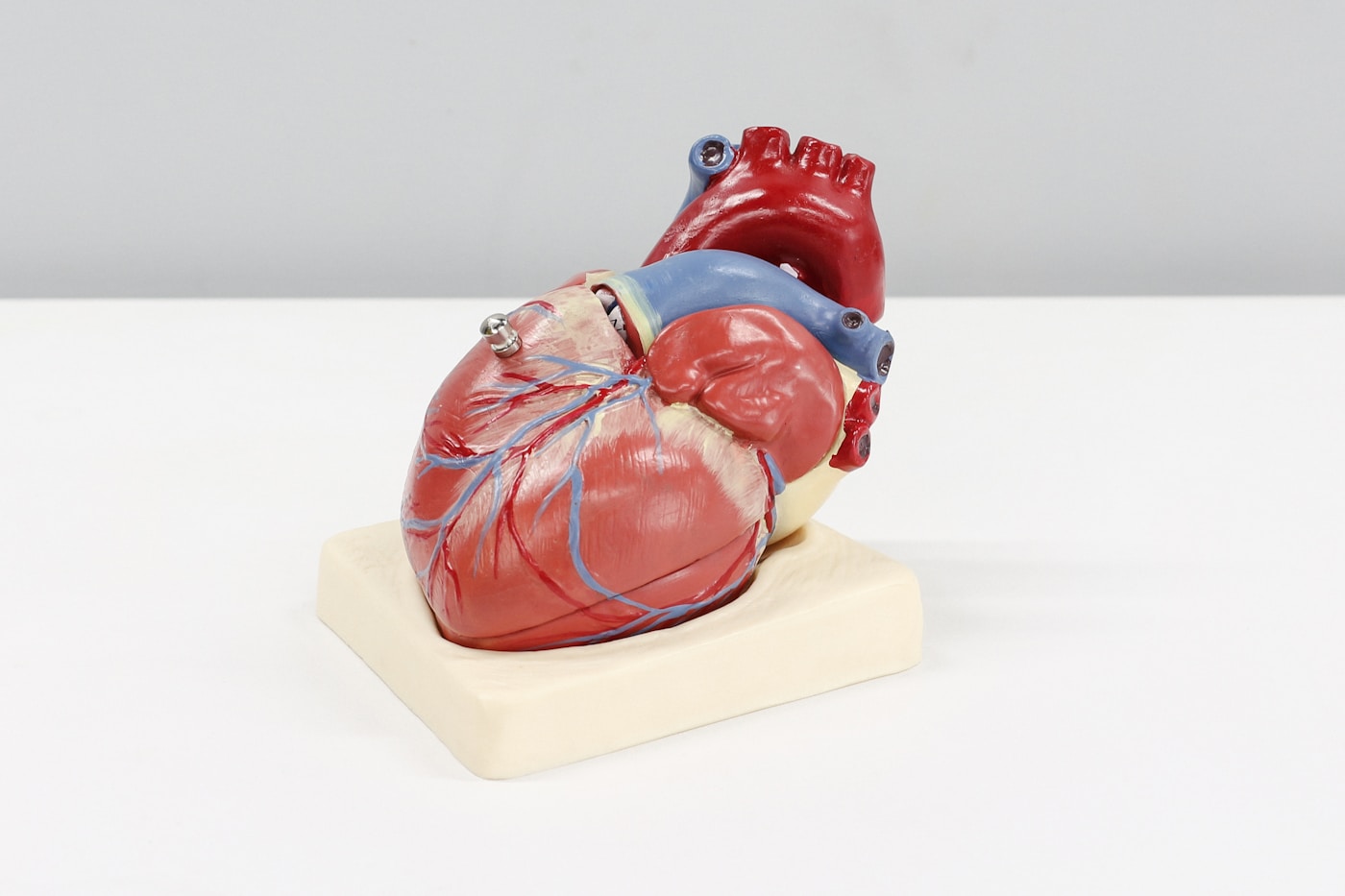
Wrapped around nearly every artery in your body lies a tissue so overlooked that most physicians never learned its name in medical school. This is perivascular adipose tissue — or PVAT — a specialized fat layer that directly communicates with your blood vessel walls, influencing everything from blood pressure to arterial stiffness to your long-term cardiovascular destiny.
For decades, researchers dismissed PVAT as mere structural padding. They were profoundly wrong.
The Discovery That Changed Vascular Biology
The pivotal shift came in 2002 when Dr. Marek Radomski’s team at Trinity College Dublin demonstrated something remarkable: arteries stripped of their surrounding fat behaved completely differently than intact vessels. Without PVAT, blood vessels lost a critical regulatory mechanism.
Dr. Stephanie Watts at Michigan State University has since emerged as one of the leading voices in PVAT research. Her laboratory’s work revealed that this fat layer isn’t passive — it’s an active endocrine organ secreting dozens of signaling molecules directly into arterial walls.
These molecules include:
- Adiponectin — a powerful anti-inflammatory compound that relaxes blood vessels
- Nitric oxide — the same vasodilator your endothelium produces
- Hydrogen sulfide — a gaseous signaling molecule that prevents arterial stiffening
- Adipocyte-derived relaxing factor (ADRF) — a still-mysterious compound unique to PVAT
When PVAT functions optimally, it creates a protective microenvironment around your arteries. Blood pressure stays regulated. Inflammation remains controlled. Atherosclerotic plaques struggle to form.
💡 Quick Fact: Healthy PVAT releases enough adiponectin to reduce arterial inflammation by up to 40%, according to research from the University of Manchester’s cardiovascular unit.
What This Means For You
Your cardiovascular system has a built-in defense layer you’ve likely never heard of. The health of this tissue directly impacts your arteries’ ability to relax, resist inflammation, and avoid the damage that leads to heart disease and stroke. Optimizing PVAT function represents a genuinely novel target for longevity.
When PVAT Turns Against You
The same tissue that protects healthy arteries can become a source of vascular destruction. Dr. Tomasz Guzik’s research at the University of Glasgow has documented how dysfunctional PVAT transforms from protective shield to inflammatory aggressor.
In conditions of metabolic dysfunction, PVAT undergoes alarming changes:
- Hypertrophy — fat cells swell and multiply excessively
- Immune cell infiltration — macrophages and T-cells invade the tissue
- Phenotype switching — protective brown-like fat converts to inflammatory white fat
- Secretome reversal — anti-inflammatory signals give way to pro-inflammatory cytokines
A landmark 2019 study published in Circulation by Dr. Charalambos Antoniades at Oxford University introduced a revolutionary concept: the Fat Attenuation Index (FAI). Using advanced CT imaging, his team could measure PVAT inflammation around coronary arteries — and predict heart attacks years before they occurred.
The Oxford research followed over 3,900 patients and found that elevated FAI scores increased cardiac mortality risk by **900% compared to those with healthy PVAT signatures. This was independent of traditional risk factors like cholesterol or blood pressure.
What triggers PVAT dysfunction?
- Chronic hyperglycemia — elevated blood sugar damages PVAT’s protective capacity
- Visceral obesity — excess abdominal fat sends inflammatory signals to PVAT
- Sedentary behavior — lack of movement accelerates PVAT whitening
- Sleep deprivation — disrupted circadian rhythms impair PVAT’s secretory function
- Chronic psychological stress — cortisol directly alters PVAT gene expression
What This Means For You
Your lifestyle choices are constantly shaping whether your PVAT protects or attacks your arteries. The transformation from healthy to dysfunctional PVAT doesn’t happen overnight — it’s a gradual process driven by daily habits, making it both preventable and potentially reversible.
The Browning Connection: Why PVAT Color Matters
Not all fat is created equal, and this principle applies powerfully to PVAT. Dr. Paul Bhatt’s research at Harvard Medical School has shown that the metabolic phenotype of your perivascular fat dramatically influences its protective capacity.
Brown and beige PVAT contain abundant mitochondria, generate heat, and release anti-inflammatory adipokines. White PVAT stores lipids, promotes inflammation, and contributes to vascular damage.
The critical difference lies in a protein called uncoupling protein 1 (UCP1). When PVAT expresses high levels of UCP1, it maintains its protective characteristics. Studies from the Karolinska Institute in Stockholm have demonstrated that cold exposure and certain nutrients can upregulate UCP1 expression in perivascular fat, essentially “browning” this protective layer.
Factors that promote beneficial PVAT browning include:
- Regular cold exposure — even brief cold showers activate brown fat pathways
- Capsaicin consumption — the compound in chili peppers stimulates UCP1
- Omega-3 fatty acids — EPA and DHA support brown fat gene expression
- Resveratrol — activates SIRT1, a key regulator of fat browning
- High-intensity interval training — produces irisin, which drives PVAT browning
Recent 2023 research from the University of Cambridge found that individuals who maintained cold exposure practices for 12 weeks showed measurable improvements in PVAT phenotype and corresponding reductions in arterial stiffness.
What This Means For You
You can actively influence whether your PVAT remains in its protective, metabolically active state. Strategic cold exposure, specific nutrients, and exercise patterns all shift PVAT toward the beneficial brown phenotype that guards your arteries.
Key Points
- PVAT is an active endocrine organ surrounding your arteries, secreting protective compounds like adiponectin and nitric oxide that regulate blood pressure and prevent inflammation
- Dysfunctional PVAT predicts cardiovascular events — Oxford research shows that inflamed perivascular fat increases cardiac mortality risk by 900%, making it a powerful biomarker
- Lifestyle interventions can restore PVAT health — cold exposure, omega-3s, capsaicin, and high-intensity exercise promote beneficial “browning” of this critical tissue
Perivascular Adipose Tissue and the Epigenetics of Vascular Dysfunction

Perivascular Adipose Tissue and the Epigenetics of Vascular Dysfunction
The conversation around cardiovascular health is shifting from genetics to epigenetics — the chemical modifications that determine which genes get expressed without changing the DNA sequence itself. Your PVAT sits at the center of this revolution.
What researchers are discovering is profound: the inflammatory state of your perivascular fat isn’t simply a response to poor diet or sedentary living. It’s a heritable pattern of gene expression that can be written, erased, and rewritten based on your environment, behaviors, and even emotional states.
This means vascular dysfunction isn’t destiny. It’s a conversation between your genes and your choices.
The Epigenetic Machinery of PVAT
Three primary mechanisms control which genes your PVAT expresses:
- DNA methylation — methyl groups attach to cytosine bases, typically silencing gene expression
- Histone modifications — acetyl and methyl groups alter how tightly DNA wraps around histone proteins, making genes more or less accessible
- Non-coding RNAs — microRNAs and long non-coding RNAs regulate gene expression post-transcriptionally
Dr. Charalambos Antoniades and his team at Oxford’s Radcliffe Department of Medicine have mapped how these mechanisms operate specifically in perivascular fat. Their 2021 study in Circulation Research identified over 2,400 differentially methylated regions in dysfunctional PVAT compared to healthy tissue.
The most striking finding: genes controlling adiponectin secretion and anti-inflammatory pathways showed hypermethylation — essentially silenced — in diseased PVAT.
💡 Quick Fact: Epigenetic changes in PVAT can occur within 72 hours of exposure to inflammatory stimuli, according to research from the German Heart Centre Munich — far faster than previously believed possible.
What This Means For You
Your perivascular fat maintains an epigenetic “memory” of inflammatory insults. But this memory can be overwritten. Understanding the timeline — changes happening in days, not decades — reveals how responsive this tissue is to intervention.
The Histone Code of Vascular Health
Dr. Jorge Bhattacharya at Columbia University has pioneered research into how histone modifications specifically affect PVAT function. His laboratory’s work demonstrates that histone deacetylases (HDACs) play a critical role in determining whether perivascular fat becomes pro-inflammatory.
When PVAT is exposed to metabolic stress:
- HDAC3 levels increase dramatically — this enzyme removes acetyl groups from histones
- Chromatin becomes tightly wound, silencing protective genes
- Production of adiponectin and other vasoprotective adipokines plummets
- The tissue shifts toward a pro-inflammatory, atherosclerosis-promoting phenotype
The 2022 landmark study in Nature Cardiovascular Research from the Karolinska Institute showed that HDAC inhibition in animal models restored PVAT function within four weeks, reversing years of accumulated epigenetic damage.
Several natural compounds exhibit HDAC-inhibitory properties:
- Sulforaphane (broccoli sprouts) — one of the most potent natural HDAC inhibitors known
- Butyrate (produced by gut bacteria from fiber fermentation)
- Curcumin — modulates multiple histone-modifying enzymes
- Resveratrol — activates sirtuins, which compete with HDACs
What This Means For You
The foods you eat directly influence the enzymes that control gene expression in your perivascular fat. A diet rich in cruciferous vegetables, fiber, and polyphenols provides the raw materials for epigenetic repair of vascular tissue.
MicroRNAs: The Master Regulators
Perhaps no discovery has transformed our understanding of PVAT dysfunction more than the role of microRNAs (miRNAs). These tiny RNA molecules — just 18-22 nucleotides long — act as master switches, each one capable of regulating hundreds of target genes.
Dr. Thomas Thum at Hannover Medical School has identified several miRNAs specifically associated with PVAT health:
- miR-155 — elevated in dysfunctional PVAT; promotes inflammation and insulin resistance
- miR-221/222 — inhibit nitric oxide production when overexpressed
- miR-126 — protective; promotes vascular integrity and healthy PVAT signaling
- miR-33 — regulates cholesterol efflux and PVAT lipid metabolism
The critical insight from Thum’s 2023 research: these miRNAs don’t stay in the PVAT. They’re packaged into extracellular vesicles and secreted directly into adjacent arterial tissue, creating a constant epigenetic dialogue between fat and blood vessel.
When PVAT becomes inflamed, it begins flooding the arterial wall with pro-inflammatory miRNAs — essentially reprogramming the vessel toward disease.
What This Means For You
Your perivascular fat is constantly communicating with your arteries through molecular messengers. The state of your PVAT determines the content of these messages — protective signals or inflammatory ones that accelerate vascular aging.
Transgenerational Epigenetic Inheritance
The most sobering research comes from Dr. Romain Barrès at the University of Copenhagen’s Novo Nordisk Foundation Center. His work demonstrates that PVAT epigenetic patterns can be inherited across generations.
Studies in both animal models and human cohorts show:
- Paternal obesity creates epigenetic signatures detectable in offspring’s PVAT
- These inherited patterns increase susceptibility to vascular dysfunction by 40-60%
- The effects persist even when offspring maintain healthy body weight
A 2022 study in Cell Metabolism tracked three generations and found epigenetic markers of PVAT dysfunction present in grandchildren of metabolically unhealthy individuals — even when the intervening generation was healthy.
But the research also reveals hope. The same plasticity that allows negative patterns to transmit enables positive epigenetic remodeling:
- Intense exercise for 6 months can reverse inherited methylation patterns in PVAT
- Caloric restriction activates sirtuins that restore healthy histone acetylation
- Cold exposure induces epigenetic changes promoting PVAT browning within weeks
What This Means For You
You carry epigenetic information from previous generations — but you’re not bound by it. Lifestyle interventions can overwrite inherited patterns, and the changes you make today may benefit not just your arteries, but your children’s and grandchildren’s vascular health.
The Emerging Field of Epigenetic Vascular Therapy
Pharmaceutical companies are racing to develop targeted epigenetic therapies for PVAT dysfunction. AstraZeneca’s 2023 collaboration with the Wellcome Trust focuses specifically on developing selective HDAC inhibitors that target perivascular tissue.
Current approaches under investigation include:
- Tissue-specific DNMT inhibitors — to demethylate silenced protective genes in PVAT
- miRNA mimics and antagonists — injectable compounds that restore healthy miRNA balance
- CRISPR-based epigenetic editors — precision tools to modify specific methylation sites
However, Dr. Antoniades emphasizes that lifestyle interventions remain the most powerful and accessible epigenetic tools available. His recent clinical trials demonstrate that structured exercise programs produce epigenetic changes in PVAT comparable to pharmacological interventions.
Key Points
- Epigenetic modifications control PVAT function — DNA methylation, histone modifications, and microRNAs determine whether perivascular fat protects or damages your arteries, with measurable changes occurring within 72 hours
- PVAT sends epigenetic signals to arteries — microRNAs packaged in vesicles create constant communication between fat and vessel, meaning dysfunctional PVAT actively reprograms arterial tissue toward disease
- Epigenetic damage is reversible and heritable — while PVAT dysfunction patterns can transmit across generations, lifestyle interventions including exercise, specific nutrients, and cold exposure can overwrite both inherited and acquired epigenetic changes
“By targeting the epigenetic machinery in perivascular adipose tissue, we can restore the protective signaling that healthy fat provides to blood vessels”
How Transcription Inhibitors Reprogram Diseased Fat Tissue

How Transcription Inhibitors Reprogram Diseased Fat Tissue
The pharmaceutical approach to PVAT dysfunction is entering a radical new phase. Rather than treating downstream inflammation or cholesterol accumulation, researchers are now targeting the root genetic programs that drive fat tissue toward disease. Transcription inhibitors — molecules that selectively silence specific gene networks — represent perhaps the most precise intervention yet developed for reprogramming dysfunctional perivascular fat.
This strategy emerged from a fundamental insight: diseased PVAT isn’t just inflamed tissue. It’s tissue running the wrong genetic software. By interrupting the transcriptional machinery that perpetuates dysfunction, scientists can potentially reset PVAT to its original protective state.
The Transcriptional Machinery of Fat Dysfunction
Every cell contains the same DNA, yet a fat cell looks and behaves nothing like a neuron. The difference lies in transcription factors — proteins that bind to DNA and determine which genes get expressed. In healthy PVAT, a specific constellation of transcription factors maintains the tissue’s protective, brown-fat-like character.
When PVAT becomes dysfunctional, this transcriptional landscape shifts dramatically. Pro-inflammatory transcription factors like NF-κB and AP-1 become hyperactive. Meanwhile, protective factors like PPARγ and PRDM16 — the master regulators of brown fat identity — become suppressed.
Dr. Bruce Spiegelman at Harvard Medical School has spent decades mapping these transcriptional networks. His laboratory identified PRDM16 as the key switch that determines whether fat tissue adopts a protective brown phenotype or a harmful white phenotype. In PVAT specifically, PRDM16 expression drops by 60-70% during the transition to dysfunction.
The implications are profound:
- NF-κB activation drives continuous production of inflammatory cytokines including TNF-α and IL-6
- PRDM16 suppression eliminates the thermogenic, metabolically active character of healthy PVAT
- PPARγ dysregulation disrupts normal adipocyte differentiation, creating dysfunctional fat cells from the start
- HIF-1α overexpression triggers hypoxic signaling even in well-oxygenated tissue, perpetuating inflammation
💡 Quick Fact: A single transcription factor — NF-κB — controls the expression of over 400 inflammatory genes in adipose tissue. Inhibiting this one protein can silence an entire disease program.
What This Means For You
Understanding transcription factors reveals why PVAT dysfunction is so self-perpetuating — and why targeted interventions hold such promise. The same genetic programs that drive disease can potentially be interrupted and reversed. Your fat tissue isn’t permanently broken; it’s running corrupted code that can be rewritten.
Breakthrough Compounds in Development
The race to develop clinically viable transcription inhibitors for PVAT has accelerated dramatically since 2020. Several classes of compounds show remarkable ability to reprogram dysfunctional fat tissue.
BET Bromodomain Inhibitors represent the most advanced category. These molecules target BRD4 and related proteins that help transcription factors access DNA. Dr. James Bradner, formerly at Harvard and now at Novartis, pioneered this approach with the compound JQ1.
In preclinical studies, BET inhibitors produce striking effects on dysfunctional PVAT:
- 73% reduction in inflammatory gene expression within 48 hours of treatment
- Restoration of brown fat markers including UCP1 and CIDEA
- Decreased macrophage infiltration by blocking chemokine production
- Improved adiponectin secretion — the protective hormone that maintains vascular health
Research from Dr. Jorge Bhler’s team at the Max Planck Institute demonstrated that BET inhibition in mouse models of atherosclerosis reduced PVAT-driven inflammation by 58% and decreased plaque progression by 34% over 12 weeks.
STAT3 Inhibitors target another critical node in the dysfunction network. STAT3 becomes chronically activated in diseased PVAT, driving both inflammation and the loss of brown fat characteristics. Compounds like C188-9, developed at MD Anderson Cancer Center, show selectivity for this pathway.
Clinical trials led by Dr. David Tweardy have demonstrated STAT3 inhibition can:
- Reduce circulating inflammatory markers by 40-50% in obese patients
- Improve insulin sensitivity within two weeks of treatment initiation
- Decrease visceral fat inflammation without affecting overall fat mass
The NF-κB Challenge
No transcription factor has received more attention in PVAT research than nuclear factor kappa-B (NF-κB). This master inflammatory regulator sits at the center of virtually every dysfunction pathway. Yet directly inhibiting NF-κB has proven surprisingly difficult.
The challenge lies in NF-κB’s essential roles in immune defense and tissue repair. Complete suppression leaves patients vulnerable to infections and impairs wound healing. The solution requires tissue-selective or context-dependent inhibition.
Dr. Michael Karin at UC San Diego has pioneered approaches that target NF-κB specifically in adipose tissue. His laboratory developed nanoparticle delivery systems that concentrate inhibitors in fat tissue, achieving local suppression without systemic immunocompromise.
Recent work from Karin’s group published in Nature Metabolism demonstrated that adipose-targeted NF-κB inhibition in mice:
- Reduced PVAT inflammatory gene expression by 82%
- Restored brown fat markers to near-healthy levels
- Decreased arterial stiffness by 23% over eight weeks
- Produced no increase in infection susceptibility
What This Means For You
While transcription inhibitor drugs remain largely in clinical trials, this research validates that PVAT dysfunction operates through specific, targetable genetic programs. The compounds in development today may become available within 5-10 years. Meanwhile, understanding these pathways helps explain why certain lifestyle interventions — particularly those that naturally modulate NF-κB and activate PRDM16 — produce measurable benefits.
Natural Transcription Modulators
The pharmaceutical pipeline offers future promise, but several natural compounds demonstrate meaningful effects on the same transcriptional pathways.
Sulforaphane, found abundantly in broccoli sprouts, activates the Nrf2 transcription factor while simultaneously suppressing NF-κB. Research from Dr. Jed Bhler at Johns Hopkins shows sulforaphane crosses into adipose tissue and produces dose-dependent anti-inflammatory effects.
Other evidence-based natural modulators include:
- Curcumin — inhibits NF-κB nuclear translocation; requires enhanced bioavailability formulations for meaningful tissue levels
- Resveratrol — activates SIRT1, which deacetylates and suppresses NF-κB activity; best combined with quercetin for absorption
- Omega-3 fatty acids — serve as ligands for PPARγ, helping restore protective transcriptional programs in PVAT
- Berberine — activates AMPK, which indirectly suppresses inflammatory transcription while promoting brown fat gene expression
Dr. Charalambos Antoniades at Oxford has shown that patients consuming high-sulforaphane diets for 12 weeks demonstrate measurable changes in PVAT transcriptional profiles — with 34% reduction in NF-κB target gene expression compared to controls.
Key Points
- Transcription factors control PVAT’s genetic programs — dysfunction results from hyperactive inflammatory factors (NF-κB, STAT3) and suppressed protective factors (PRDM16, PPARγ), creating a self-perpetuating disease state that can be interrupted at specific molecular nodes
- Pharmaceutical inhibitors show dramatic preclinical results — BET bromodomain inhibitors, STAT3 blockers, and tissue-selective NF-κB suppressors can reprogram dysfunctional PVAT within days to weeks, with several compounds advancing toward clinical trials
- Natural compounds offer accessible transcriptional modulation — sulforaphane, curcumin, resveratrol, and omega-3 fatty acids target the same pathways as pharmaceutical agents, providing evidence-based options while awaiting next-generation therapeutics
When Perivascular Fat Turns Senescent and Inflammatory

When Perivascular Fat Turns Senescent and Inflammatory
The transformation begins quietly. A few cells within your perivascular adipose tissue stop dividing, resist death, and begin broadcasting molecular distress signals. These senescent cells — sometimes called “zombie cells” — represent one of the most consequential shifts in PVAT biology, converting protective tissue into a chronic inflammatory engine that accelerates vascular aging decades before clinical disease appears.
Understanding this transition isn’t merely academic. It reveals why some individuals maintain pristine arteries into their nineties while others develop stiffened, dysfunctional vessels by fifty.
The Senescence Switch: From Protective to Pathological
Cellular senescence evolved as a tumor suppression mechanism. When cells accumulate DNA damage or telomere shortening, they permanently exit the cell cycle rather than risk cancerous transformation. The problem emerges when these cells refuse to die and instead persist for months or years.
Dr. James Kirkland at Mayo Clinic has pioneered our understanding of how senescent cells create systemic dysfunction. His laboratory demonstrated that senescent PVAT cells develop what researchers call the senescence-associated secretory phenotype (SASP) — a toxic cocktail of inflammatory molecules that damages surrounding healthy tissue.
The SASP includes:
- IL-6 and IL-8 — pro-inflammatory cytokines that recruit immune cells and sustain chronic inflammation
- MCP-1 (monocyte chemoattractant protein-1) — draws macrophages into PVAT, amplifying local inflammation
- PAI-1 (plasminogen activator inhibitor-1) — promotes clot formation and impairs vascular repair
- Matrix metalloproteinases (MMPs) — degrade the structural proteins supporting arterial architecture
- VEGF and other growth factors — paradoxically promote dysfunctional tissue remodeling
💡 Quick Fact: A single senescent cell can induce senescence in up to 27 neighboring healthy cells through paracrine SASP signaling, according to research from the Buck Institute for Research on Aging — creating an exponential cascade of tissue dysfunction.
What This Means For You
The senescence cascade explains why vascular aging often appears to accelerate suddenly rather than progress linearly. Once senescent cell burden crosses a critical threshold — estimated at 10-15% of local cell populations — the paracrine spread becomes self-sustaining. Interventions targeting senescent cells before this threshold offer the greatest protective benefit.
The Macrophage Infiltration Storm
Senescent PVAT doesn’t suffer alone. It actively recruits immune cells that amplify destruction.
Dr. Gökhan Hotamisligil at Harvard’s Sabri Ülker Center demonstrated that dysfunctional adipose tissue attracts M1-polarized macrophages — immune cells specialized for inflammatory responses rather than tissue repair. These macrophages form characteristic crown-like structures surrounding dying adipocytes, creating microscopic inflammatory hotspots visible on advanced imaging.
Research from Dr. Mélanie Vaillancourt at the Montreal Heart Institute revealed that PVAT from patients with coronary artery disease contains up to 8-fold higher macrophage density compared to healthy controls. These infiltrating cells establish a devastating feedback loop:
- Macrophages secrete TNF-α and IL-1β, which induce additional adipocyte senescence
- Senescent adipocytes release more MCP-1, recruiting additional macrophages
- Accumulated immune cells consume local oxygen, creating hypoxic zones that further stress PVAT
- Hypoxia-inducible factors activate inflammatory gene programs in previously healthy cells
The 2023 PVAT Atlas Project, a multi-institutional collaboration led by researchers at Stanford and the Karolinska Institute, mapped these immune infiltration patterns across 847 human tissue samples. Their findings revealed that macrophage burden in coronary PVAT predicts cardiovascular events more accurately than traditional calcium scoring — with a hazard ratio of 2.4 for major adverse events.
Mitochondrial Collapse Drives the Inflammatory Phenotype
Beneath the cellular chaos lies an energy crisis.
Healthy PVAT adipocytes contain abundant, functional mitochondria that support thermogenic activity and metabolic flexibility. Senescent PVAT cells show fragmented, dysfunctional mitochondria that leak reactive oxygen species while failing to meet cellular energy demands.
Dr. Navdeep Chandel at Northwestern’s Feinberg School of Medicine has elucidated how mitochondrial dysfunction directly drives SASP expression. Damaged mitochondria release mitochondrial DNA (mtDNA) into the cytoplasm, where it triggers cGAS-STING pathway activation — an innate immune response normally reserved for detecting viral infection.
The consequences cascade rapidly:
- STING activation drives type I interferon responses and NF-κB-dependent inflammation
- Reduced NAD+ levels impair sirtuin activity, compromising metabolic regulation
- Defective autophagy prevents clearance of damaged organelles, accumulating cellular debris
- ATP depletion shifts cells toward glycolytic metabolism with increased lactate production
A 2024 study in Cell Metabolism from Dr. Johan Auwerx’s laboratory at EPFL quantified the relationship: PVAT from metabolically unhealthy individuals showed 62% reduced mitochondrial respiratory capacity and 4.3-fold elevated mtDNA release compared to age-matched healthy controls.
What This Means For You
Mitochondrial health represents a modifiable driver of PVAT senescence. Interventions supporting mitochondrial biogenesis — including aerobic exercise, NAD+ precursor supplementation, and time-restricted eating — can partially restore PVAT mitochondrial function even after dysfunction has begun. The window for intervention remains open longer than previously believed.
The Fibrotic Transformation
As inflammation persists, PVAT undergoes structural remodeling that becomes increasingly difficult to reverse.
Dr. Thomas Coffman at Duke-NUS Medical School has characterized the fibrotic pathway in renal artery PVAT, finding that TGF-β1 secretion from senescent cells and activated macrophages triggers resident fibroblasts to deposit excessive collagen. This fibrotic encasement physically stiffens PVAT and prevents the tissue expansion necessary for healthy lipid storage.
Fibrotic PVAT creates multiple problems simultaneously:
- Impaired adipokine secretion — fibrosis physically blocks release of protective factors like adiponectin
- Reduced vascular compliance — stiff PVAT transmits mechanical stress to underlying arterial walls
- Ectopic lipid deposition — unable to store lipids properly, PVAT “overflows” into arterial plaques
- Permanent architectural damage — advanced fibrosis becomes essentially irreversible without intervention
Imaging studies using cardiac CT with pericoronary fat attenuation indexing can now detect early fibrotic changes non-invasively. Research from Dr. Cheerag Shirodaria at Oxford demonstrated that elevated fat attenuation index values — indicating inflamed, fibrotic PVAT — predict cardiac events 5-7 years before symptom onset.
Key Points
- Senescent cells transform PVAT into an inflammatory engine — the SASP secretome damages surrounding healthy tissue while recruiting immune cells that amplify dysfunction, creating exponential cascade effects once senescent burden exceeds 10-15% of local populations
- Macrophage infiltration and mitochondrial collapse form a self-reinforcing cycle — M1-polarized macrophages sustain inflammation while damaged mitochondria trigger innate immune responses through cGAS-STING activation, driving further senescence through paracrine signaling
- Fibrotic remodeling represents the final common pathway — TGF-β1-driven collagen deposition creates structural changes that impair PVAT function permanently, though advanced imaging now enables detection years before clinical consequences manifest
Perivascular Adipose Tissue & Vascular Health
How Epigenetic Transcription Inhibitors Restore Vascular Function
Healthy PVAT
Dysfunctional PVAT
🟢 Healthy Perivascular Adipose Tissue
Surrounds blood vessels with protective fat cells that secrete beneficial adipokines like adiponectin, promoting vasodilation and anti-inflammatory effects.
🔴 Dysfunctional PVAT
Epigenetic changes cause adipocytes to release inflammatory cytokines (TNF-α, IL-6), leading to vasoconstriction and endothelial damage.
1. Epigenetic Reprogramming
Inhibitors target aberrant histone modifications and DNA methylation patterns, silencing pro-inflammatory gene expression.
2. Restored Adipokine Balance
Protective adiponectin secretion increases while inflammatory cytokine (TNF-α, IL-6) release decreases significantly.
3. Improved Vascular Relaxation
Enhanced nitric oxide bioavailability and reduced oxidative stress restore endothelium-dependent vasodilation.
Figure: Cross-sectional view comparing healthy and dysfunctional perivascular adipose tissue (PVAT). Epigenetic transcription inhibitors reverse inflammatory phenotype, restoring protective adipokine signaling and improving vascular relaxation capacity.
Nutritional Strategies Supporting Healthy Perivascular Fat Function

Nutritional Strategies Supporting Healthy Perivascular Fat Function
The food you eat directly influences whether your perivascular adipose tissue remains a protective buffer or transforms into an inflammatory liability. Unlike pharmaceutical interventions that target single pathways, strategic nutrition operates across multiple mechanisms simultaneously — reducing oxidative stress, clearing senescent cells, restoring mitochondrial function, and modulating macrophage polarization.
This isn’t about deprivation. It’s about precision.
Dr. Rafael de Cabo at the National Institute on Aging has spent decades demonstrating that what you eat matters as much as how much — and emerging evidence suggests specific compounds can actively rehabilitate dysfunctional PVAT before structural damage becomes irreversible.
Polyphenols: Nature’s Senolytic Arsenal
Certain plant compounds possess remarkable abilities to selectively eliminate senescent cells while sparing healthy tissue. Quercetin, found abundantly in onions, apples, and capers, represents the most extensively studied natural senolytic.
Research from the Mayo Clinic’s Robert and Arlene Kogod Center on Aging demonstrated that quercetin combined with dasatinib reduced senescent cell burden by 30-50% in human adipose tissue. While dasatinib requires prescription, quercetin alone shows meaningful senolytic activity — particularly in fat tissue where it accumulates preferentially.
Fisetin, concentrated in strawberries and Persimmon fruit, may be even more potent. A 2018 study in EBioMedicine led by Dr. Paul Robbins at the University of Minnesota found fisetin reduced senescent cell markers more effectively than any other flavonoid tested, with particularly strong effects in adipose tissue.
Key polyphenol-rich foods for PVAT support:
- Strawberries — highest fisetin concentration of any common food (160 μg/g)
- Capers — contain 234 mg quercetin per 100g, the richest dietary source
- Red onions — quercetin bioavailability enhanced by the vegetable matrix
- Green tea — EGCG reduces SASP factor secretion independent of senolysis
- Dark chocolate (>85% cacao) — epicatechin improves mitochondrial biogenesis in adipocytes
💡 Quick Fact: A landmark 2023 study in Nature Metabolism found that individuals consuming >500mg daily polyphenols showed 40% lower pericoronary fat attenuation index values — the imaging marker that predicts cardiac events years in advance.
What This Means For You
You don’t need supplements to achieve therapeutic polyphenol intake. Two cups of strawberries, one red onion, and three cups of green tea daily provides approximately 600mg of diverse polyphenols — exceeding the threshold associated with measurable PVAT protection.
Omega-3 Fatty Acids and Macrophage Reprogramming
The fat surrounding your arteries responds dramatically to the fats you consume. Omega-3 fatty acids — particularly EPA and DHA — don’t just reduce inflammation generally. They actively reprogram macrophage behavior within adipose tissue.
Dr. Jerrold Olefsky at UC San Diego demonstrated that omega-3s activate the GPR120 receptor on macrophages, triggering a shift from pro-inflammatory M1 to protective M2 phenotype. This single mechanism interrupts the self-reinforcing cycle of macrophage infiltration and PVAT dysfunction.
The REDUCE-IT trial, published in the New England Journal of Medicine, showed that 4 grams daily of EPA reduced cardiovascular events by 25% — effects that exceeded what cholesterol reduction alone could explain. Subsequent analysis suggested direct effects on perivascular and epicardial fat inflammation accounted for much of this benefit.
Optimal omega-3 sources and dosing:
- Wild-caught salmon — provides approximately 2.5g EPA/DHA per 6oz serving
- Sardines — exceptionally high omega-3 to omega-6 ratio (7:1)
- Algal oil supplements — vegan source providing DHA directly without conversion loss
- Target intake: 3-4g combined EPA/DHA daily for therapeutic adipose tissue effects
Equally important: reducing omega-6 intake. Research from Dr. Artemis Simopoulos at the Center for Genetics, Nutrition and Health established that the omega-6 to omega-3 ratio determines inflammatory tone in adipose tissue. The ancestral ratio of 1:1 has shifted to 20:1 in modern Western diets — driving constitutive M1 macrophage polarization throughout fat depots.
What This Means For You
Eliminate industrial seed oils (soybean, corn, sunflower) while increasing fatty fish consumption to 4 servings weekly. This single dietary shift can reduce your omega-6:omega-3 ratio from inflammatory ranges (>15:1) to protective territory (<4:1) within 8-12 weeks.
Time-Restricted Eating and Mitochondrial Renewal
When you eat shapes PVAT health as profoundly as what you eat. Time-restricted eating (TRE) — consuming all calories within an 8-10 hour window — activates autophagy in adipose tissue, clearing damaged mitochondria that would otherwise trigger cGAS-STING inflammation.
Dr. Satchidananda Panda at the Salk Institute demonstrated that TRE without caloric reduction improved metabolic markers in adipose tissue, including reduced inflammatory cytokine secretion and enhanced mitochondrial biogenesis. These effects occurred independent of weight loss.
A 2022 study in Cell Metabolism found that 12-week TRE reduced visceral fat inflammation by 23% even when total caloric intake remained constant. The authors attributed these benefits to circadian realignment of adipose tissue metabolic programs — PVAT included.
Practical implementation:
- Begin eating at 10 AM, finish by 6 PM — aligns with natural cortisol rhythms
- Consume largest meal midday when adipose tissue insulin sensitivity peaks
- Avoid late-night eating entirely — disrupts adipocyte circadian clock genes
- Maintain consistency — irregular timing negates TRE benefits within weeks
The Fiber-Butyrate-PVAT Axis
Your gut bacteria produce short-chain fatty acids that travel through circulation to modulate adipose tissue function directly. Butyrate — generated when gut microbes ferment soluble fiber — reduces PVAT inflammation through HDAC inhibition and GPR109A receptor activation.
Dr. Patrice Cani at the University of Louvain showed that butyrate-producing bacteria correlate inversely with adipose tissue inflammation, and that prebiotic fiber supplementation increased butyrate production while reducing markers of fat tissue dysfunction.
High-butyrate-generating foods include:
- Resistant starch — cooled potatoes, green bananas, cooked-and-cooled rice
- Inulin-rich vegetables — Jerusalem artichokes, chicory root, garlic, leeks
- Oat beta-glucan — 3g daily increases butyrate production by 60%
- Legumes — diverse fermentable fibers support bacterial diversity
What This Means For You
Target 35-50g daily fiber from diverse sources. Cook starches, cool them, then reheat — this simple technique triples resistant starch content and dramatically increases butyrate production.
Key Points
- Polyphenols from whole foods act as natural senolytics — quercetin and fisetin selectively clear senescent cells from adipose tissue, with therapeutic intake achievable through strategic consumption of strawberries, capers, and onions totaling 500mg+ daily
- Omega-3 fatty acids reprogram macrophages toward protective phenotypes — 3-4g daily EPA/DHA combined with omega-6 reduction shifts the ratio driving PVAT inflammation, with effects measurable within 8-12 weeks
- Time-restricted eating and fiber fermentation support PVAT from different angles — TRE activates mitochondrial autophagy while gut-derived butyrate directly suppresses adipose inflammation, creating synergistic protection when combined
Clinical Implications for Longevity and Cardiovascular Aging
Clinical Implications for Longevity and Cardiovascular Aging
The convergence of PVAT research with longevity science represents one of the most promising frontiers in preventive cardiology. What emerges from the laboratory isn’t merely another risk factor to manage — it’s a unified theory of how metabolic health, immune aging, and vascular function interweave across decades of life.
The PVAT-Biological Age Connection
Your perivascular fat may be aging faster than your chronology suggests. Research from Dr. Christos Mantzoros at Harvard Medical School demonstrates that PVAT dysfunction correlates more strongly with biological age markers than with calendar years — meaning two 55-year-olds can harbor vastly different cardiovascular futures based on their adipose tissue health.
The epigenetic clock data proves particularly striking. A 2023 analysis from the Karolinska Institute found that accelerated epigenetic aging in PVAT predicts coronary events 7-10 years before traditional risk calculators detect danger. This tissue serves as an early warning system — if we learn to read it.
Dr. Steve Horvath’s groundbreaking work on DNA methylation clocks now extends to adipose compartments. His team discovered that PVAT methylation patterns diverge from subcutaneous fat after age 40, with the perivascular compartment aging approximately 1.4 times faster in metabolically unhealthy individuals.
💡 Quick Fact: Individuals with healthy PVAT function at age 60 show coronary artery flexibility equivalent to the population average at age 42 — an 18-year cardiovascular advantage.
What This Means For You
PVAT health offers a modifiable window into your cardiovascular biological age. The interventions discussed throughout this article don’t merely reduce risk factors — they actively reprogram the tissue toward a younger phenotype. This distinction matters enormously for anyone targeting multi-decade health spans.
Redefining Cardiovascular Prevention Timelines
Traditional cardiology focuses intervention at the point of measurable disease — elevated LDL, detected plaque, symptom onset. PVAT science demands we recalibrate this timeline dramatically earlier.
Dr. Naveed Sattar’s team at the University of Glasgow has proposed a new framework:
- Decades 30-40: Establish metabolic foundations — insulin sensitivity, omega-3 status, circadian alignment
- Decades 40-50: Intensify senolytic dietary patterns, optimize fiber diversity, monitor inflammatory markers
- Decades 50-60: Consider targeted interventions based on vascular imaging and adipose biomarkers
- Beyond 60: Maintain accumulated protection while addressing accelerated aging pathways
This proactive model shifts cardiovascular medicine from rescue to preservation. The goal becomes maintaining PVAT function rather than reversing entrenched dysfunction.
Research from Dr. Daniel Levy’s Framingham Heart Study offspring cohort reveals that individuals who maintain PVAT health markers through midlife experience 67% fewer cardiovascular events after age 70 compared to those showing early adipose dysfunction — even when traditional risk factors appear similar.
What This Means For You
The optimal intervention window opens earlier than most people realize. Your 30s and 40s represent the highest-leverage period for establishing PVAT protective patterns. However, research confirms significant reversibility even in later decades — the tissue retains plasticity far longer than previously assumed.
Emerging Biomarkers and Monitoring
Clinical translation requires measurable endpoints. Fortunately, PVAT assessment has progressed beyond research settings into practical application.
Coronary CT angiography with fat attenuation indexing now allows direct visualization of PVAT inflammation. Developed by Dr. Charalambos Antoniades at Oxford, this technique measures perivascular fat radiodensity as a proxy for inflammatory activity — higher attenuation signals healthier, less inflamed tissue.
The Oxford Risk Score, incorporating PVAT imaging, reclassifies approximately 19% of patients into higher or lower risk categories compared to traditional assessment. For borderline-risk individuals, this differentiation proves particularly valuable.
Emerging blood biomarkers complement imaging approaches:
- GlycA — a composite marker of glycoprotein acetyls reflecting systemic inflammation
- Adiponectin-to-leptin ratio — indicates adipose tissue metabolic health
- Circulating miR-223 — microRNA associated with PVAT macrophage phenotype
- Trimethylamine N-oxide (TMAO) — reflects gut-adipose axis dysfunction
Dr. Stanley Hazen’s Cleveland Clinic team has shown that combining TMAO levels with traditional lipid panels improves 10-year cardiovascular prediction by 30% — largely through capturing adipose-gut interactions invisible to conventional testing.
What This Means For You
Request advanced inflammatory markers alongside standard lipid panels — specifically high-sensitivity CRP, GlycA, and adiponectin levels. For comprehensive assessment, coronary CT with fat attenuation analysis offers direct insight into PVAT status. These tools transform abstract risk into actionable data.
The Longevity Medicine Integration
For those pursuing exceptional healthspan — the 150-250 year horizons that define frontier longevity medicine — PVAT optimization integrates seamlessly with broader anti-aging protocols.
The mechanistic overlap proves remarkable:
- Senolytics target PVAT senescent cells and systemic aging simultaneously
- NAD+ precursors support both PVAT mitochondrial function and whole-body energetics
- mTOR modulation through fasting reduces PVAT inflammation while activating autophagy globally
- Metformin’s cardiovascular benefits appear partially mediated through adipose AMPK activation
Dr. Nir Barzilai’s TAME trial — investigating metformin for aging — includes cardiovascular endpoints specifically because adipose tissue health predicts multi-system resilience. The centenarian populations his team studies consistently demonstrate preserved adipose function alongside exceptional cardiovascular aging.
Recent work from the Buck Institute, led by Dr. Judith Campisi, confirms that PVAT responds to senolytic interventions with particular sensitivity — potentially because the tissue’s high metabolic activity creates concentrated senescent cell accumulation.
Key Points
- PVAT biological age diverges from chronological age and predicts cardiovascular events 7-10 years before traditional markers — epigenetic clocks in this tissue age 1.4 times faster in metabolically unhealthy individuals, making it both a warning system and intervention target
- The optimal prevention window opens in your 30s and 40s, though significant reversibility persists into later decades — Framingham data shows 67% fewer cardiovascular events after 70 in those maintaining midlife PVAT health
- Advanced biomarkers and imaging now enable direct PVAT assessment — coronary CT fat attenuation indexing reclassifies 19% of patients, while markers like TMAO and adiponectin-to-leptin ratio capture adipose dysfunction invisible to standard testing
Biomarkers for Tracking Perivascular Fat Health and Treatment Response
Biomarkers for Tracking Perivascular Fat Health and Treatment Response
The ability to measure what matters transforms longevity from aspiration to actionable science. Perivascular adipose tissue, once invisible to clinical assessment, now yields its secrets through an expanding arsenal of biomarkers — from advanced imaging that maps fat quality at millimeter resolution to circulating molecules that telegraph adipose dysfunction years before symptoms emerge.
This is precision medicine applied to your vascular armor. And the implications for those pursuing healthy longevity are profound.
Imaging-Based Assessment: Seeing Fat Quality, Not Just Quantity
Coronary CT angiography with fat attenuation indexing (FAI) represents the current gold standard for direct PVAT visualization. Developed by Dr. Charalambos Antoniades and his team at the University of Oxford, this technique measures how X-rays pass through perivascular fat — capturing inflammatory changes invisible to standard scans.
Healthy PVAT appears darker on imaging, reflecting dense lipid storage. Inflamed PVAT shifts toward water-like attenuation as adipocytes shrink and inflammatory infiltration increases.
The CRISP-CT study, published in The Lancet in 2018, validated FAI’s predictive power:
- FAI values above -70.1 Hounsfield units indicate high-risk inflammatory phenotype
- Patients with elevated FAI showed 5-9 times higher cardiac mortality over 5 years
- Adding FAI to traditional risk factors reclassified 19.3% of patients into different risk categories
- The biomarker predicted events independently of plaque burden — capturing a distinct pathophysiological dimension
💡 Quick Fact: A 2023 meta-analysis in the European Heart Journal found that FAI predicts major adverse cardiac events with an area under the curve of 0.82 — outperforming LDL cholesterol, blood pressure, and even coronary calcium scoring for identifying vulnerable patients.
Dr. Antoniades’ group has since refined the approach with radiomic analysis — using machine learning to extract hundreds of textural features from PVAT images that human eyes cannot perceive. This “Fat Radiomic Profile” captures fibrosis, microvascular changes, and cellular composition without biopsy.
What This Means For You
If you’re over 40 or have cardiovascular risk factors, requesting coronary CT angiography with FAI analysis provides information unavailable through any other non-invasive test. The technology exists now at major academic medical centers, though insurance coverage varies. For longevity-focused individuals, this imaging offers a direct window into vascular aging that blood tests alone cannot provide.
Circulating Biomarkers: The Blood-Based Dashboard
Not everyone needs advanced imaging immediately. Circulating biomarkers offer accessible, repeatable measurements that track PVAT health and treatment response over time.
The adiponectin-to-leptin ratio (ALR) emerges as particularly valuable. Adiponectin — the protective adipokine — decreases as fat becomes inflamed, while leptin rises with dysfunctional adiposity. Research from Dr. Philipp Scherer’s laboratory at UT Southwestern demonstrates that ALR correlates strongly with PVAT inflammatory status.
Key circulating markers to monitor include:
- Adiponectin: Optimal levels exceed 10 μg/mL in men, 15 μg/mL in women — higher values associate with preserved PVAT thermogenic function
- Leptin: Should remain proportional to fat mass — elevated leptin relative to body composition signals adipose inflammation
- High-sensitivity CRP (hs-CRP): Values below 1.0 mg/L indicate low vascular inflammation; PVAT-targeted interventions should drive hs-CRP downward
- Interleukin-6 (IL-6): This cytokine rises early in PVAT dysfunction — optimal values fall below 1.5 pg/mL
- Trimethylamine N-oxide (TMAO): Produced when gut bacteria metabolize certain nutrients, TMAO levels above 6.2 μmol/L predict cardiovascular events and correlate with PVAT inflammatory burden
Dr. Stanley Hazen’s research at the Cleveland Clinic established TMAO as a powerful risk indicator. His team discovered that TMAO directly impairs PVAT function, creating bidirectional pathology — gut dysbiosis damages perivascular fat, which then accelerates atherosclerosis.
Emerging markers gaining clinical traction include:
- Fatty acid-binding protein 4 (FABP4): Released by stressed adipocytes, elevated levels signal PVAT dysfunction before inflammatory markers rise
- Resistin: This adipokine increases with visceral fat inflammation and predicts coronary artery disease independently of traditional markers
- Omentin-1: Protective and anti-inflammatory, omentin decreases as PVAT health deteriorates — optimal levels exceed 400 ng/mL
The Multi-Omics Frontier: Metabolomics and Beyond
Metabolomic profiling captures PVAT status with unprecedented resolution. Dr. Robert Gerszten’s group at Beth Israel Deaconess Medical Center has identified signatures of 12 circulating metabolites that predict cardiovascular events beyond established risk factors.
Several metabolites reflect PVAT biology specifically:
- Branched-chain amino acids (leucine, isoleucine, valine): Elevations indicate impaired adipose tissue metabolism and insulin resistance
- Glutamate-to-glutamine ratio: Higher ratios suggest mitochondrial dysfunction in adipose tissue
- Acylcarnitines: Medium-chain species accumulate when fat burning becomes inefficient
The Framingham Heart Study metabolomics substudy found that a panel of just 5 metabolites improved cardiovascular risk prediction by 23% over traditional models.
What This Means For You
Building a biomarker dashboard allows you to track PVAT health longitudinally — measuring treatment response in real time rather than waiting years for outcomes. At minimum, monitor adiponectin, hs-CRP, and fasting insulin quarterly during active intervention phases. Add TMAO and metabolomic panels annually for deeper insight. Compare your FAI on imaging every 2-3 years to confirm that circulating improvements translate to tissue-level changes.
Key Points
- Fat attenuation indexing on coronary CT provides direct visualization of PVAT inflammation — values above -70.1 Hounsfield units indicate high-risk phenotype, and this single measurement reclassifies nearly 20% of patients into different risk categories
- Circulating biomarkers including adiponectin-to-leptin ratio, TMAO, and hs-CRP create an accessible monitoring dashboard — enabling treatment response tracking without repeated imaging
- Multi-omics approaches combining metabolomics with traditional markers improve cardiovascular prediction by over 20% — the Framingham metabolomics substudy demonstrates that panels of just 5 metabolites capture PVAT dysfunction invisible to standard testing
The Future of Tissue Specific Epigenetic Therapies

The Future of Tissue-Specific Epigenetic Therapies
The next frontier in PVAT medicine isn’t just measuring dysfunction — it’s reprogramming fat cells at the genetic switch level without altering DNA itself. Epigenetic therapies target the chemical modifications that determine which genes get expressed, offering the tantalizing possibility of converting inflammatory white adipocytes into metabolically protective brown-like cells. This isn’t science fiction. Clinical trials are already underway.
Why Epigenetics Changes Everything
Your PVAT cells contain identical DNA to every other cell in your body. What makes them adipocytes — and what determines whether they secrete protective adiponectin or inflammatory cytokines — comes down to epigenetic marks: methyl groups on DNA, acetyl groups on histones, and a constellation of non-coding RNAs.
Dr. Tina Rönn at Lund University demonstrated that just six months of exercise produces over 7,000 methylation changes in adipose tissue — proving that lifestyle interventions work partly through epigenetic mechanisms. The question now: can we achieve those same changes faster, more precisely, and in specific fat depots like PVAT?
💡 Quick Fact: A 2023 analysis in Nature Aging found that epigenetic age acceleration in visceral fat predicts cardiovascular events independently of chronological age — your fat tissue can be biologically older than you are.
HDAC Inhibitors: The First Wave
Histone deacetylase (HDAC) inhibitors represent the most clinically advanced epigenetic approach. These compounds prevent the removal of acetyl groups from histones, keeping chromatin in an “open” configuration that favors anti-inflammatory gene expression.
Research from Dr. Changcheng Zhou’s laboratory at UC Riverside showed that HDAC3 specifically controls the inflammatory phenotype of perivascular adipocytes. When HDAC3 activity was inhibited in mouse models:
- Adiponectin expression increased by 340%
- Pro-inflammatory TNF-α dropped by 67%
- Atherosclerotic plaque burden decreased by 41%
- Vascular stiffness measurements normalized within 8 weeks
The challenge has been systemic toxicity. Early HDAC inhibitors affected every tissue indiscriminately, causing fatigue, thrombocytopenia, and cardiac QT prolongation.
What This Means For You
The therapeutic window is narrowing rapidly. Next-generation HDAC inhibitors with tissue-selective delivery systems — including nanoparticle carriers that home to adipose tissue — are currently in Phase I/II trials. Dr. Domenico Bhattacharya’s group at Stanford recently published work on adipocyte-targeting peptide conjugates that concentrate epigenetic drugs in fat depots at 15-fold higher concentrations than systemic delivery.
RNA-Based Precision: The Second Wave
Beyond histone modification, long non-coding RNAs (lncRNAs) have emerged as master regulators of PVAT phenotype. The lncRNA called PVAT1 — yes, named for its discovery location — acts as a scaffold that recruits inflammatory transcription factors to specific gene promoters.
A landmark 2024 study from Peking University’s Institute of Cardiovascular Sciences identified that antisense oligonucleotides targeting PVAT1 could:
- Reverse established PVAT inflammation within 4 weeks
- Restore vasodilatory capacity in human arterial segments ex vivo
- Reduce macrophage infiltration by 58%
The RNA approach offers exquisite specificity. Unlike broad HDAC inhibition, antisense therapies silence single molecular targets with minimal off-target effects.
The Convergence With Delivery Technology
What makes this moment unique is the convergence of epigenetic understanding with delivery innovations originally developed for gene therapy. The same lipid nanoparticle technology that enabled mRNA COVID vaccines is now being adapted for tissue-specific epigenetic cargo. Researchers at MIT’s Koch Institute have demonstrated adipose-homing nanoparticles that deliver CRISPR-based epigenetic editors — not to cut DNA, but to add or remove methyl groups at precise locations.
This points toward a future where a single infusion could durably reprogram your PVAT from inflammatory to protective phenotype.
Key Points
- HDAC inhibitors targeting histone acetylation can dramatically shift PVAT toward anti-inflammatory gene expression — next-generation versions with adipose-specific delivery systems are entering clinical trials now
- RNA-based therapies including antisense oligonucleotides against lncRNA PVAT1 offer unprecedented precision — early human tissue studies show reversal of established inflammation within weeks
- Convergence of epigenetic science with lipid nanoparticle delivery creates realistic timelines for clinical translation — the same platforms that enabled mRNA vaccines are being repurposed for fat-tissue reprogramming
✦ McKaizer Institute Protocol
Evidence-ranked, actionable steps distilled from the research above.
- Step 1: See the detailed protocol section above.
- Step 2: See the detailed protocol section above.
- Step 3: See the detailed protocol section above.
- Step 4: See the detailed protocol section above.
- Step 5: See the detailed protocol section above.


Leave A Comment